Showing posts with label protein. Show all posts

Wednesday, April 10, 2024

Designing a new antibiotic to combat drug resistance


In this article by Sharon Reynolds, the author discusses the development of a new antibiotic, cresomycin, that has been effective in treating drug-resistant infections in mice.

Antibiotics typically bind to and shut down bacterial ribosomes, the organelle that builds proteins in the cell. Due to antibiotic resistance, bacteria have evolved mechanisms to prevent the blocking of their ribosomes by antibiotics. Researchers have developed a fully synthetic compound - cresomycin - based on the molecular structure of lincosamide antibiotics. They tested this compound in mice with antibiotic-resistant E. coli and found that it suppressed bacterial growth. When tested in human cells, the compound didn't cause notable damage. Researchers are hoping to move closer to clinical trials next.

I found this to be a very informative article. Antibiotic resistance is a very serious problem, so discovering the development of potential new antibiotics is incredibly important. I do think the article was purposefully vague when discussing the damage the antibiotic did to human cells in testing. What precisely does "notable damage" mean? Regardless, this is undoubtedly a step in the right direction. 


Article: Designing a new antibiotic to combat drug resistance

Additional Information: Bringing multidrug-resistant pathogens to their knees

Friday, November 18, 2022

Fluorescent Mouse Blood Helping to Find Brain Diseases

Albumin Protein


Scientists conducting a study with mice at the University of Copenhagen have discovered a way to make the mice's blood fluorescent so that it can be seen as it moves through the body and brain. The process works with a protein produced in the liver called albumin. The researchers took a gene on a fluorescent protein and attached it to a gene of albumin. The modified albumin is then inserted into a genetically modified virus and the virus is injected into a mouse's tail, which has large blood vessels. The virus causes the mice no harm but enters the liver and tricks it into making the modified albumin which makes the blood fluoresce. As the blood moves throughout the body and goes into places like the brain, the flow of blood can be studied and analyzed to find out more about diseases like Alzheimer's, depression, and even strokes. 

The new fluorescent blood method is a game changer for these types of studies because it lasts in the animal for months. Before this method, the main way to highlight blood and trace its flow was by chemical dyes and this lasted for only hours. Now blood can be traced over much longer periods of time and be used to trace long-term disease progression. The method is also being applauded by advocates for the ethical use of animals in research. The method is less painful and stressful for mice since it only requires one injection whereas the dye method required many reinjections since the dye disappeared over a few hours and had to be reinserted. 

I think this was very fascinating research and really showed how advancements in genetics have allowed for this great new type of technology to be developed. This new method of using fluorescent blood is a revolutionary development and I believe will be very helpful to researchers and tracing blood flow for years to come. Being less stressful for lab animals is another plus of this study and I feel is another important advantage of this new method. 

Monday, November 14, 2022

Genetically Engineered Plants Produce More Vegetable Oil

Scientists in Singapore have published their research on genetically altering the plant protein WRINKLED1 (WRI1) to produce more vegetable oil. The Nanyang Technological University in Singapore conducted the analysis, experimenting with the plant Arabidopsis, a member of the mustard family. The experimental results showed that by modifying the WRI1 protein Arabidopsis could produce 15-18% more oil in its seeds. The breakthrough for the research team came when they were able to complete a high-resolution image of the structure of WRI1. The completed structure revealed how the protein binds to DNA and this tells the plant how much oil to make in seeds. To do this the scientists modified the crystal structure of the WRI1 to increase the amount of binding to DNA that takes place. "The stronger the binding the more oil the plant will concentrate in its seeds" was the idea of the study said Asst Prof Ma, a plant molecular biologist in the study. Currently, the team has filed for a patent for their gene modification technique and is looking for partners to commercialize the new technique. 

I found this research to be very interesting. We have discussed a lot about the genetics of domestic crops in class so far, and I think that this is the next big step forward to ensuring crops can provide enough food for the population. I think that genetically modifying crops will become more and more prevalent in the future 
as studies like these continue to make advances in altering the genetic makeup of crops to make them produce more, grow bigger, be more tolerant to pests or droughts, and have other traits of interest. The applications of this field are practically limitless and it will be interesting to see where else this and other studies will take it. 

Thursday, September 15, 2022

Researchers found a species of Jellyfish that are effectively immortal


In an article written by author Veronique Greenwood, she talks about how a small species of jellyfish known as Turritopsis dohrnii, that although these jellyfish swim and eat like many other jellyfish, they have a secret that sets them apart from other sea creatures. Turritopsis dohrnii live in the Mediterranean Sea and are secretly known to turn their old age back into their young selves. For Turritopsis dohrnii, when their bodies get damaged, the mature medusas can turn back time and transform themselves to their immature life stages. To do this, T. dohrnill sheds its limbs, allowing them to drift around in the water column and morph into a polyp form, which then allows them to attach themselves to rocks, plants or other sea life. After researching the jellyfish’s genome to search for the gene that controls this unusual life cycle, the only scientist, Shin Kubota at Kyoto University in Japan, found, after sequencing the genome, is that the jellyfish had extra copies of a particular gene. These additional genes allowed the researchers to see that these genes protect and repair the jellyfish’s DNA, which is an essential aspect of their survival. To test this rejuvenation process, the researchers put Turritopsis dohrnii under stressful conditions by not feeding them. The adult medusas shrank into smaller sizes, sprouted into polyps, and began remaking their adult bodies. During this process, the scientists captured snapshots of what genes the jellyfish were using in each stage of their development. In each phase, the scientists took jellyfish and froze them to turn them into mush to extract their mRNA, which gave them records of which genes were being used to make proteins. The scientists found that in the adults, their genes were relatively active, meaning they were used more frequently to make proteins. And as the jellyfish descended into the polyp phases, their genes became quieter, making less protein to form those smaller sizes.


I personally think that this type of science is so fascinating and could possibly help for future research for humans and for other living species. If jellyfish and humans have the same genes this could lead to a new era in medicine. Since researchers were able to figure out which genes were being used for each life stage, this could be a glimpse into future research of possible cancer treatments or other treatments of human health and medicine. 

Friday, December 10, 2021

Archaea Microbes can Morph Their DNA


Single-celled archaea microbes can expand and stretch their DNA to get easy access to their genetic material. Researchers studied the organism Methanothermus fervidus, and saw that the DNA was constantly in motion. While complex multicellular organisms rely on molecular machinery to regular gene expression, Achaea microbes can contort their DNA to regulate expression. Cyro-electron microscopy shows that microbes can pack their chromatic into tight coils that can spring open with unexpected contortions. Researchers stated that the structure looks very similar to slinkies. It is hypothesized that the microbes have this slinky structure to bend genes so they can turn it on and off. This is in contrast to other cellular organisms that might use chaperone proteins and other machinery to turn genes off or on. It is still being studied if their silky-like DNA can maintain that shape in solution. Karoline Luger stated that "studying strange organisms like archaea can help scientists answer questions about multicellular life."

Thursday, December 9, 2021

The Genes that are Behind Cat's Spots & Stripes

Cats have spots and stripes all over their body on their fur. Do you ever wonder how it gets there? Researchers have found a specific gene that all domestic and wild big cat species develop spots and stripes. The gene that was responsible for this is the DKK4 gene which is found to be active in thick skin as opposed to thinner skin. It was found to be known for fur with a blur of colors which is a mutation from the DKK4 gene. The all-white and all-black cats have patterns underneath their fur. However, the pigment lacks in white cats as opposed to other solid-colored cats because the pattern is overridden by instructions to produce one color. The WNTs protein is also essential because it works closely with DKK4 to form a prepattern that is 2 to 3 millimeters long. The color differences that control the coat color variation are also similar to the gene that's responsible in cheetahs and thicker, more prominent fur patterns. Other than the WNTs protein and DDK gene, there are more unknown genes that are behind why some cats have spots and why some cats have stripes. 

Thursday, November 25, 2021

You are Getting Sleepy: Tiredness, Proteins, and DNA Repair


Researchers in Israel have been working to unravel the mystery of sleep by examining the mechanisms that cause organisms to get tired. Homeostatic sleep pressure builds up in our body the longer we stay awake and begins to decrease as we fall asleep. But what causes homeostatic pressure to increase to the point of tiredness and how does sleep cause it decrease? The pressure is built during waking hours due to accumulating DNA damage in neurons, caused by things like UV light, neuronal activity, oxidative stress, radiation, and enzymatic errors. And during sleep, sleep recruit repair systems correct DNA breaks that cause excessive DNA damage in our brains. To see if DNA damage was the trigger for homeostatic pressure and sleep state, the researchers examined zebrafish. These fish are particularly easy to study because of their nocturnal sleeping patterns and simple brain that is comparable to a human's. 

Methods involving irradiation, pharmacology, and optogenetics were used to induce DNA damage in the zebrafish. They discovered that after subjecting the zebrafish to light interruption during the dark period when they'd be sleeping, six hours of sleep was enough to reduce DNA damage. But with less than six hours of sleep, DNA damage was not adequately reduced and the zebrafish continued to sleep during daylight hours. The mechanism that tells them (and us) to sleep is the protein PARP1. It's the among the first to rapidly respond to DNA damage sites in cells and recruits relevant systems to clean out the damage. PARP1 increasingly clusters at DNA break sites during wakefulness and decreases during sleep. Inhibition of the protein in zebrafish meant that the fish weren't aware they were tired, didn't sleep, and the DNA didn't get repaired. Understanding the purpose of sleep on a cellular level may help with future research on sleep disturbances, aging, and neurogenerative disorders


https://www.sciencedaily.com/releases/2021/11/211118203657.htm

Saturday, October 23, 2021

The Friend Gene


    Well, not quite what the title entails but very similar. Researchers did a study using mice, to try and find a correlation between "friend groups" and individuals that get along well with each other. In this experiment researcher, Dr. Kelly concluded that there is a biomarker present in mice that attracts them to each other. The unaltered mice with "normal" behavior and the matching biomarkers seemed to pool together and separate themselves from the altered mice. The altered mice had undergone damage to the brain's hippocampus leading to antisocial behavior. The anti-social mice also seemed to prefer interacting with similarly anti-social mice. Dr. Kelly recalled a time where researchers observed that in humans, people with autism seemed to gravitate toward others with similar conditions as opposed to unaffected individuals. During her further research with the mice, she was able to find the specific protein marker, PDE11. In the anti-social mice, they were missing the protein marker altogether. In the normal mice, the protein marker was hidden but present. Dr. Kelly believes that if we can solve this puzzle, we may be able to better match people as partners and decrease the divorce rate as well as increase the health of the general public. People can be surrounded by others who make them feel good. I found this information very interesting. I would really like to see how it works in people. I think people have more "ins and outs" per se than mice and would wonder if it would still, despite the complexity of human beings', work?

https://www.sciencedaily.com/releases/2021/09/210901090115.htmhttps://www.sciencedaily.com/releases/2021/09/210901090115.htm

https://pubmed.ncbi.nlm.nih.gov/14574133/

https://www.medschool.umaryland.edu/news/2021/Do-Genetics-Control-Who-Our-Friends-Are-It-Seems-So-with-Mice.html

Friday, April 23, 2021

The Protein Siglec-XII May Be Contributing to Humans' High Cancer Rates

According to an article written by Asher Jones from The Scientist, there is a protein that may be contributing to the high rates of cancer in humans. This protein is called the Siglec-XII which is possibly promoting carcinoma progression in humans. These proteins are found on innate immune cells and involved in cell signaling. Siglec-XII is encoded by the SIGLEC12 gene and in this when it is no longer binding with sialic acid, it starts to have abnormal cell signaling. Nissi Varki and Ajit Varki, researchers from UC San Diego of Medicine had stated that protein plays a role in the progression of cancer and could explain why the rate of cancer progression is high in humans. 


The researchers had found that about 30% of people that produce the Siglec-XII protein are more than twice the risk of developing a more advanced cancer compared to people who do not produce the protein. The protein is expressed on epithelial cells and has unique human features such as a fixed homozygous missense mutation which inactivates its natural ligand recognition property, and a polymorphic frameshift mutation that eliminates the full-length protein expression in approximately 60-70% of worldwide human populations, etc.  It is quite remarkable that researchers are discovering how cancer cells are being promoted and I think if they are able to figure out exactly how this is happening then they might be able to find a way to stop the protein from forming and may be able to reduce the rate of cancer in humans. 


Monday, April 12, 2021

A Genetic Patch that Eliminates Hearing Problems associated with the DFNA9 Gene


According to the researchers at the Radboud University, a genetic patch can eliminate hearing problems associated with the DFNA9 gene. Usually, the mutation in the DFNA9 gene causes deafness from birth. In some cases, hearing problems can occur after forty years. If inherited a mutated copy of the gene, then hearing problems will develop later in life. If inherited two normal copies of the gene, then that person will have normal hearing. The genetic patch, known as antisense oligonucleotide, works by developing a small piece of RNA that binds to the RNA messenger from the muted DFNA9 gene. This stops the production of the mutant DFNA9 protein. Adding the genetic patch would prevent further damage to the ear. Another option to treat problems associated with the DFNA9 gene is to turn it off. This would also stop the production of the mutant DFNA9 protein.  


Articles:

https://neurosciencenews.com/genetic-patch-deafness-17935/

https://www.cell.com/molecular-therapy-family/nucleic-acids/pdf/S2162-2531(21)00068-8.pdf?_returnURL=https%3A%2F%2Flinkinghub.elsevier.com%2Fretrieve%2Fpii%2FS2162253121000688%3Fshowall%3Dtrue

Friday, April 9, 2021

Have this protein, run the risk of getting Cancer by 2x


There is a protein that is possibly running the risk of getting cancer by doubling it. A Singlec, which is a cell surface group of proteins, that are sialic-acid binding immunoglobulins, are involved in cell signaling in immune cells. The gene SIGLEC12 which encodes for Siglec-XII is now not binding to sialic acid and has been found to abnormally cell signaling in humans. This was discovered by Ajit & Nissi Varki, who work at UC San Diego School of Medicine. They believe this is linked to people having a great chance at carcinoma cancer progression where SInglec-XII is found in abundance. For the most part, people have a mutation that inactivates the SIGLEC12 gene altogether, but there are some that don’t put them at higher risk. It was also seen that people with this gene, have a worse late prognosis if the cancer is not caught early on. Specifically, on patients with colorectal cancer, it was discovered that around 80% of patients had the operational form of SIGLEC12.

Further research has been done on mice. They introduced tumor cells, that is to produce the SIGLEC12 gene. When monitored in comparison to tumors that do not have the SINGLEC12 gene, it was discovered to see a growth rate that was doubled.

Now, this is very interesting. Since we see this gene playing a big role in certain cancers in the way they form. We could also try to be able to catch this early on patients. Therefore, hopefully, it will lead to more success of remission since it progresses rapidly. But of course, the problem is, genetic testing is not widely available yet, but I hope one day to see it a part of a doctor's daily routine check-up. Our genetics tell us what our body is made of and if we could catch the anomalies before they eve occur, these would be the advances I like to see.

https://health.ucsd.edu/news/releases/Pages/2020-12-08-evolution-may-be-to-blame-for-high-risk-of-advanced-cancers-in-humans.aspx

https://www.the-scientist.com/the-literature/rogue-protein-could-contribute-to-humans-high-cancer-rates-68576

https://www.technologynetworks.com/cancer-research/news/amp/protein-manipulates-metabolism-to-block-cancer-cell-death-347187

Monday, November 9, 2020

Gene editing may help obesity in future


Humans have different types of fats. One type is white type which is what makes human obese. Another type is the brown type which burns energy and keeps people lean. Scientists in this article have tried to treat obesity by turning this white fat into brown. To achieve this, the researchers used CRISPR-Cas9. They used this technology to insert a molecular switch into the DNA of white fat cells. This boosted how much protein was made, which then turned the white fat cells into human brown-like ones. This was then transplanted into mice. They did a study with many different groups of mice and measured many different numbers like blood sugar. The mice with more brown cells ended up being less obese. This scientist hopes this will one day treat obese in humans. In my opinion, I believe this is a steppingstone to curing obesity. Changing these cells to burn energy rather than store can help a lot of people. This is a good start to help genetics in humans.


Extra information

Sunday, October 18, 2020

 Common Cold Cure Possible?

Common Cold: Symptoms, Treatment, Causes, vs Flu, Duration & Prevention


    Studies completed by researchers at Stanford University and the University of California- San Francisco found it may be possible to protect ourselves from the common cold and other viral diseases by disabling just one protein in our cells. The approach of targeting proteins in cells has worked before to stop other viruses associated with asthma and polio. At least half of the common cold cases are a Rhinovirus, which are mutation prone and you can get them often. The way the researchers believe will stop this is by disabling the protein that allows for Enteroviruses, including the Rhinovirus to replicate. To see what proteins in human cells help replicate the virus. the scientists generate a. cultured line of human cells that can possibly be affected by this. When finding the cell they affect them with two different viruses and notice some cells managed to survive the infection and find the gene that had been knocked out. It managed to work on mice so maybe one day they will be able to not just protect us from the common cold but nearly all enteroviruses.

https://med.stanford.edu/news/all-news/2019/09/in-human-cells-and-mice-a-cure-for-the-common-cold.html

Tuesday, November 26, 2019

Making a Vaccine for Ebola


Director of the Center for Microbial Pathogenesis at Georgia State University, Christopher Basler, has been working with other researchers to change the genome of the Ebola virus. They managed to modify the VP35 protein in the virus, allowing it to be combated by the early immune response in animals. In doing this, they made a strain of Ebola that is safe for researchers to work with. When given to monkeys, the altered strain provoked an immune response when the monkeys were exposed to the wildtype Ebola.

Basler intends to further his research to eventually develop a drug that can modify the VP35 protein in the same was as he did in the lab, or even use the strain of Ebola to create a vaccine to prevent future cases.


Article: https://www.usnews.com/news/health-news/articles/2019-09-18/tiny-genetic-tweak-may-stop-ebola-virus-in-its-tracks

Related Article: https://news.gsu.edu/2019/09/17/biomedical-sciences-researchers-find-building-mutations-into-ebola-virus-protein-disrupts-ability-to-cause-disease-and-activates-protective-immunity/

Sunday, November 24, 2019

Microbe "Eats" Electrons

Some phototrophic microbes can pull electrons in from an electrode source and use them as fuel. They do this by using a unique processing step to regulate the production of an electron transfer protein used during this process. This process is involves getting electrons across the barrier of the bacteria, which is difficult since the outer layer is non-conducive and impermeable to iron and/or electrodes. A strain of Rhodopseudomonas palustris TIE-1 builds a channel by processing a protein called deca-heme cytochrome c, to help electrons pass through the membrane. This process is called extracellular electron uptake, or EEU, and it is done by bacteria when nutrients are low. Researchers now understand this process better and are going to use these biological markers to identify other microbes in the wild that are able to use electrons as a fuel source. Doing so will help to further understand why this process is important in metabolic evolution and microbial ecology.

Image result for rhodopseudomonas palustris tie-1

I find it very fascinating how microbes are able to develop unique ways to survive harsh conditions. Microbes are so adaptable when it comes to change in the environment and seeing a process like this is very cool. I've never heard of a bacteria using electrons as fuel before and didn't even think of it as a possibility, so learning that this is something some can do is incredible.

Link:

Related Article:

Tuesday, November 19, 2019

Extinct Ancestor to Orangutan Opens up Doors for Further Human Evolution Research

Ancient protein sequencing has been used to determine the genetic position of the Gigantopithecus blacki. It revealed that the orangutan is its closest living relative. This is the first time that genetic information this old has been able to be used from such a warm, damp environment. This is important because primates are closely related to humans, meaning it may be possible to retrieve similar information on the evolutionary line leading to humans. We used to only be able to go back about 400,000 years, but now it may be possible to trace human evolution as far back as two million years. The sequencing of proteins from dental enamel also proved to be useful when studying lineage of species, when there is no surviving DNA. Only a few jaws, but a lot of teeth were found from this species. This protein sequencing of the enamal showed that the orangutan and Gigantopithecus blacki split up about 12 million years ago. Mass spectrometry was used to analyze these protein sequences. Because there are not a lot of fossils found from Gigantopithecus blacki, a lot of speculation surrounds what its physical characteristics may have been.

Image result for orangutan

A lot is already known about the evolution of a lot of animals and of humans, but there is still so much missing. When new fossils are found and they are sequenced to find out which species they are closest related to, a whole new door is opened. From there other lineages can be mapped out and we can get a clearer understanding of how evolution occurred. What's even more interesting about this case, is that the way these fossils were sequenced opened up new possibilities for the sequencing of future fossils from extinct human species. Now more questions about the evolution of humans can be answered.

Link:
https://www.sciencedaily.com/releases/2019/11/191113153053.htm

Related Article:
https://healthsciences.ku.dk/newsfaculty-news/2019/11/extinct-giant-ape-directly-linked-to-the-living-orangutan/

Tuesday, September 24, 2019

Small Genetic Change May Stop Ebola Virus


During this experiment, scientists used monkeys and changed a single protein in the Ebola virus called VP35, which enables the Ebola virus to block immune responses to infection.  After changing the VP35 protein, it was found that the body's immune system was activated to fight off the Ebola virus.  Not only did this change in protein help fight off the virus, but it also acted as a vaccine in a way and helped to prevent the animal from being infected. After changing this protein, the scientists presented the monkeys with the Ebola virus and the monkeys were completely protected. 


With these findings, scientists want to see if they can make a drug that can change the VP35 protein in humans in order to protect them from getting the Ebola virus.  Of course, just because this method worked in an animal does not necessarily mean that it could work in humans.  They also believe that working with this VP35 protein could also produce immune responses in other diseases as well.

Ebola is a very dangerous virus that has killed a lot of humans.  I am hopeful that this technique in changing the VP35 protein works in humans and that it can help to fight off the Ebola virus, and also prevent it from ever occurring.  Like the article said, perhaps focusing on this virus can also help to prevent or fight off other diseases as well. 

https://www.usnews.com/news/health-news/articles/2019-09-18/tiny-genetic-tweak-may-stop-ebola-virus-in-its-tracks
https://www.thoughtco.com/ebola-virus-373888
https://www.cdc.gov/vhf/ebola/about.html

Monday, December 3, 2018

Implications of Jiankui's Genetically Modified Babies


The latest news in genetics comes out of Shenzhen, China where an associate professor of bioengineering at the Southern University of Science and Technology, He Jiankui, claims that two little girls are the world’s first genetically edited newborn babies. Using CRISPR, Jiankui has modified the babies’ genes to make them resistant to infection from HIV. The father of the babies is said to be positive for HIV. As of November 27th (the date of the article), there was no data to demonstrate how this experiment took place but is said to speak on more on the topic. The University has condemned the topic and even issued a statement saying that they had no idea that the project was going on.


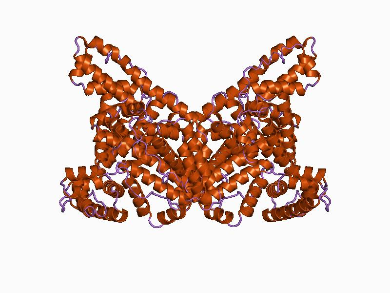
How HIV infects cells via CCR5


In a Youtube video, Jiankui claimed that he used CRISPR to disable CCR5, a protein receptor that allows HIV to infect blood cells. On specific mutation, Delta32, disables HIV from locking onto the cell. In theory, if all individuals carried this mutant allele, then nobody would be able to get AIDs from HIV. CRISPR has been used in the laboratory for many situations, like eliminating diseases and improving the health of different crops. This technique, though, has never been used on human embryos, and therefore the results are unknown. One major problem is that CRISPR can cause off-target mutations to genes away from the target genes, and therefore can have many other implications.

Many companies are already looking to gene therapies in adults to edit the CCR5 cells in adults. In theory, scientists would remove blood from HIV positive patients, delete the CCR5 protein and return the cell back to the patient. It seems like every action has a reaction, and in this sense, getting rid of the CCR5 protein would increase susceptibility to West Nile virus, which is already seen in the real world when individuals are born without the CCR5 protein. Overall, Jiankui wrote a piece that discussed the core principles in the genetic editing of human embryos.

I am very interested in ethics in relation to science, and especially in relation to genetic editing. I believe that it should be interesting to see the effects of this experiment over time. I do not feel strongly for or against human genome editing or the idea of “designer babies”. If this experiment works and is able to basically eliminate HIV and AIDs ability to infect humans, think of what other uses human genome editing could have. Genetic cancers could be cured, and other genetic diseases could be edited. I think the use of genome editing for superficial purposes is unethical at this point in time. I think that the world has a ways to go in terms of being able to accept genetic editing and there will always be disagreement for it. Overall, the effects of this experiment will be interesting to see how it changes genetics forever, and it is so cool to see this monumental moment in scientific history.

Monday, April 23, 2018

Genetic mutation causes deadly skin condition on dogs


A study has been assessed and resulted in the discovery of a deadly skin condition affecting puppies/dogs. Research reports that this discovery causes LAD, known as lethal acrodermatitis. This deadly condition causes skin lesions on the paws and face of puppies and dogs. Having such condition may delay in growth. Dogs with the condition may have a dilute, subtle coat color.

University of Bern’s Anina Bauer and her team discovered the mutation that causes lethal acrodermatitis. LAD, lethal acrodermatitis, is an inherited disease in dogs, mainly bull terriers and miniature bull terriers. This disease causes painful, infected wounds on the paws. The disease can inhibit growth or cause death before two years old. LAD in dogs can be compared to acrodermatitis enteropathica (zinc deficiency) in humans. However, unlike humans, there has been no found cure for this disease.

The mutation in the gene that codes for the protein muskelin 1 causes the affects of LAD. The mutation disallows the splicing of mRNA and leads to lack of muskelin 1. Protein muskelin 1 plays a role in shaping, as well as intracellular transport.

Research results show that scientists can only further investigate the causes of the mutation, functions of the protein muskelin 1, and perform genetic tests.

For additional information, refer to the original article.

For additional information, click the link of Anina Bauer and team’s research article and article on LAD in bull terriers.


Saturday, April 14, 2018

Cryo-electron Microscopy (Scaffolding) = Proteins in detail

Scaffolding method uses a cryo-electron microscopy finally being able to see the small proteins inside cells. Until now that method did not yield results. At the University of California Los Angeles (UCLA) a team led by Todd Yeates said, "This new method should be useful broadly". They were able to attach 12 copies of DARPin (a small protein) on a cube-shaped molecular cage, creating a larger version in a sense allowing them to view it by cryo-electron microscopy. Being able to finally visualize small proteins will help with figuring out how the disease causing ones work and "how it does what it does", said Yeates. This advancement will eventually foster new pharmaceuticals and treatments.