Showing posts with label Blood. Show all posts

Friday, November 18, 2022

Fluorescent Mouse Blood Helping to Find Brain Diseases

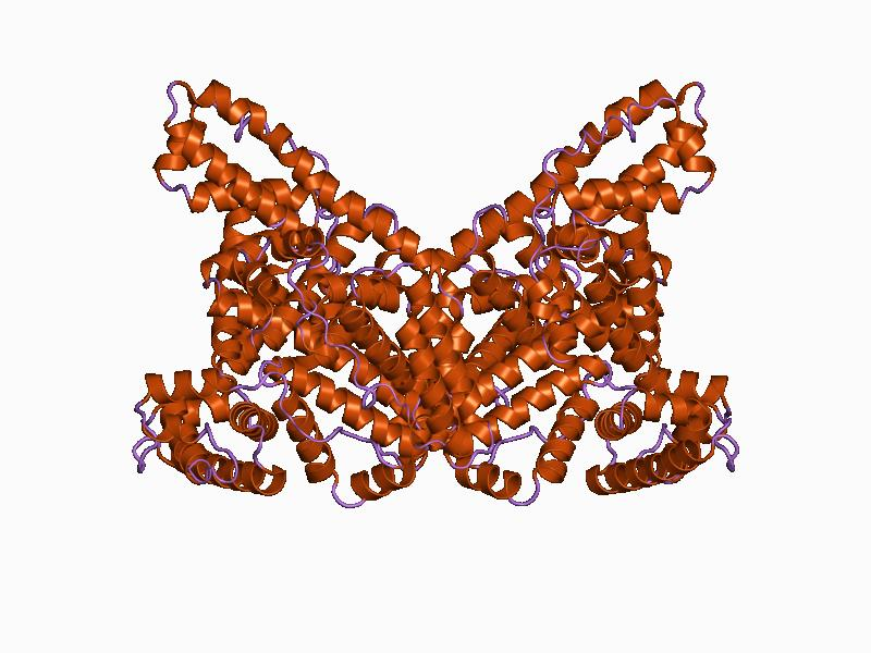
Albumin Protein


Scientists conducting a study with mice at the University of Copenhagen have discovered a way to make the mice's blood fluorescent so that it can be seen as it moves through the body and brain. The process works with a protein produced in the liver called albumin. The researchers took a gene on a fluorescent protein and attached it to a gene of albumin. The modified albumin is then inserted into a genetically modified virus and the virus is injected into a mouse's tail, which has large blood vessels. The virus causes the mice no harm but enters the liver and tricks it into making the modified albumin which makes the blood fluoresce. As the blood moves throughout the body and goes into places like the brain, the flow of blood can be studied and analyzed to find out more about diseases like Alzheimer's, depression, and even strokes. 

The new fluorescent blood method is a game changer for these types of studies because it lasts in the animal for months. Before this method, the main way to highlight blood and trace its flow was by chemical dyes and this lasted for only hours. Now blood can be traced over much longer periods of time and be used to trace long-term disease progression. The method is also being applauded by advocates for the ethical use of animals in research. The method is less painful and stressful for mice since it only requires one injection whereas the dye method required many reinjections since the dye disappeared over a few hours and had to be reinserted. 

I think this was very fascinating research and really showed how advancements in genetics have allowed for this great new type of technology to be developed. This new method of using fluorescent blood is a revolutionary development and I believe will be very helpful to researchers and tracing blood flow for years to come. Being less stressful for lab animals is another plus of this study and I feel is another important advantage of this new method. 

Monday, April 11, 2022

New Discovery Explains Vampire Bats' Thirst for Blood


An article by Scientific American explains that a new study done by scientists in Germany have developed a possible explanation as to why vampire bats drink blood. Vampire bats are an interesting subset of bats because they only feed on blood, primarily mammals. While the species thrives on livestock blood, in other species, it is considered a poor diet. 

Blood, in general, is not rich in many nutrients because it does not contain any fat or carbohydrates, and contains little calories. In order to get enough nutrients to stay alive, the bats must drink 1.4 times their body weight during a single feeding, which is 1.68 oz of blood. These levels of blood are considered dangerous because the iron levels are so high, and can be dangerous to the digestive system. Scientists wanted an answer as to how these bats only live off of blood, and if genetics possibly play a role.

Michael Hiller and his colleagues at the LOEWE Center for Translational Biodiversity Genomics in Frankfurt, Germany used vampire bat genomes to study for any possible genes (or lack thereof) to provide an explanation into this phenomenon. It turns out it is the lack of certain genes that account for their ability to only consume blood. Hiller mentions that typically, seeing a deletion or mutation in genes are areas for concern, but in this case, it is actually beneficial. 13 "key" genes have been lost and range in functions, that altogether, create the perfect recipe for the vampire bats to keep thriving on their blood diet.

The loss of two genes specifically, REP15 and CYP39A1, aid significantly in keep the bats alive and healthy. REP15 is a gene that used to help keep iron out of the bats' stomachs and transfer it into their bloodstream. With that no longer a role in the vampire bats, the iron is able to move into the intestines. However, the researchers found that this might be a good thing, as the digestive cells have a small lifespan, so they can be continually shed and replaced to help excrete the iron-rich droppings instead of allowing it to absorb in the bloodstream. CYP39A1 is another helpful loss, as it used to degrade a by-product of cholesterol digestion. This gene deletion causes the metabolite to increase heavily, and other research in rodents has shown a correlation of increased levels of the metabolite and sociability and better memory, which helps the vampire bats. All in all, this genetic research provides an answer into why this species is able to thrive on something that is not considered a nutritious food.

This article really interested me because I was not very familiar with vampire bats. I was unaware that they only drink blood and I also had no idea about the lack of nutrition that comes with consuming blood. However, the scientists' research looking into what could be responsible for allowing the bats to consume the blood was fascinating. I would have never thought that it was actually the loss of genes that helped the bats thrive over the generations. This is a very unique species with a special diet that if any other animal were to eat solely, they would die. It is interesting that genetics (or lack of) plays a crucial role in the evolution of vampire bats.

Related Articles:

Inside the Vampire Bat Diet

Additional Research Into the Loss of Genes for Vampire Bats

Wednesday, November 24, 2021

How Gene Therapy Could Combat Hemophilia

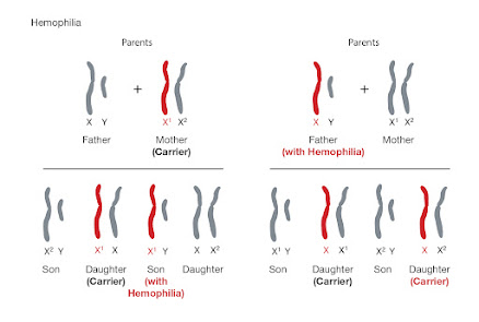
Hemophilia affects 1 in 5,000 men worldwide. It is a blood disorder in which the patient lacks the FVIII coagulation factor. This is the factor that causes blood to clot.

A new gene therapy could combat this blood disorder. The therapy, known as an adeno-associated viral (AAV) vector-based gene therapy, uses the SPK-8011 gene. This gene, often paired with steroids in the event of a negative immune response, helps the liver to express the FVIII coagulation factor in affected males. 

During the trials, researchers saw a 91.5% reduction in major or life-threatening bleeding episodes. Sixteen of the 18 patients maintained FVIII expression for their entire time in the trial. Twelve of those patients were followed for more than two years and had no apparent decrease in FVIII activity. Throughout the trial, there were no prevalent safety concerns for the patients.

More testing is needed, but if successful, this new gene therapy could be a very legitimate option for those who struggle with Hemophilia.

Thursday, July 29, 2021

Noninvasive Prenatal Testing (NIPT)


Noninvasive Prenatal Testing is a procedure used to determine the genetic code of a fertilized baby, while it is still in the first trimester. The purpose of this procedure is intended to make sure non-desirable traits are not inherited from family tree's that have a history of these traits. The procedure is done by something as simple as a blood draw. Once the blood is drawn from the maternal parent, the blood is sent to a laboratory and examined by geneticists. The results would return in 8 to 14 days. 


Link:  1https://medlineplus.gov/genetics/understanding/testing/nipt/#:~:text=Noninvasive%20prenatal%20testing%20(NIPT)%2C,in%20a%20pregnant%20woman's%20blood.

Link: https://www.mayoclinic.org/tests-procedures/noninvasive-prenatal-testing/about/pac-20384574

Sunday, October 4, 2020

What Does Your Blood Type Mean?


Hopefully we all know our blood type, I’m O negative, this is very important information if you're ever in need of a blood transfusion. But what does your blood type even mean? We generally associate blood types with three different letters A, B, O, and by four main categories, A, B, AB, O. An individual with type A blood has A antigens on the surface of their red blood cells. Their plasma contains anti-B antibodies, which would attack any B antigens attempting to enter the type A blood person. This means if someone who possesses type A blood receives type B blood during a procedure, the body would reject the blood transfusion. If you have type B blood your red blood cell surface contains B antigens and would not receive type A antigens. If someone, like myself, has type O blood, we have O antigens on our white blood cells and we have antibodies that would attack both A and B antigens. People with type O blood are considered “universal doners”. We can donate to anyone, A, B, AB and O. However individuals with type O blood can only receive type O blood. People with type AB, on the other hand, are universal receivers, they can receive A, B, AB, and O blood types. O positive is the most frequently occuring blood type and is in high demand for that reason. O negative is in the highest demand since it is universal as anyone can receive O negative blood, yet it is one of the most rare blood types.

https://www.redcrossblood.org/donate-blood/blood-types.html

https://www.medicalnewstoday.com/articles/218285#risks

Tuesday, November 19, 2019

Mutation causing Milky White Blood


Three siblings were discovered to have milky white blood due to a genetic disorder. The blood was full of fat that caused the coloration. The siblings, two of which were a set of twins, were born to a first cousin couple that are part of a Pennsylvania Dutch family. As teens the siblings experienced abdominal pain and when they sought medical help they were diagnosed with hypertriglyceridemia. This disorder causes an increase of triglycerides in the blood. However, further testing recently done in their fifties showed that they actually have an ultra rare condition known as familial chylomicronemia syndrome causing very high levels of triglycerides causing the color of the blood to turn milky white. 
Figure above shows the molecular form of a triglyceride
The siblings have long attempted to control their triglyceride levels to alleviate the symptoms of abdominal pain, fever and vomiting.  There was only one gene found to cause the condition when normally there are multiple. The gene usually breaks down triglycerides in the blood. Each sibling was found to have two copies of the mutated gene which must have been passed down from each parent. The Particular gene mutation found in the children has never been seen before. Their condition can be helped by a low fat diet.
I was intrigued by the title of this article “A Rare Genetic Disorder Turned These Siblings' Blood 'Milky' White” because I had no idea that this could occur. I found it interesting that these patients blood had so much fat in it that it became a white color. I was surprised that this condition could be alleviated by a controlled diet.


Wednesday, October 2, 2019

Convert blood types into type O


                        During the 256th National Meeting & Exposition of the American Chemical Society, researchers from the University of British Columbia describe they may have found a way to convert blood group into blood type O.  Researchers believe using a bacterial enzyme will turn any blood into type O.  The bacteria enzyme is from the human gut. To convert any blood types AB, A, B, into O, the markers or antigens from these types of blood will need to remove. By removing these antigens, all blood will be compatible. This is just a theory right now. Currently, researchers are continuing to work on this new idea. So far, they had gotten outstanding results from samples of human feces.

             This idea is very calm and astonishment. Right now, there are four basic types of blood groups. There are AB, A, B, and O. Each of these blood groups has different antigens. Blood A has A antigens and can only get it A blood, blood AB has A antigens and B antigens and can receive any type. Blood B has B antigens and can only get B blood.

On the other hand, blood O has no antigens but can give to any of the blood groups. Also, blood O can only receive from O. If in the future, this succeeds, everyone will no longer need to wait for blood donations. All blood will be compatible with each other.


https://bigthink.com/stephen-johnson/scientists-use-gut-bacteria-to-convert-any-blood-into-type-o
https://techthelead.com/scientists-discovered-how-to-convert-type-a-blood-into-type-o/

Wednesday, April 11, 2018

Leeches: Unexpected Helper to Studying Biodiversity

Leeches. The gross, worm-like vampires of the natural world that can swell to 10x their body size have recently been found to be of some great value. Dr. Michael Tessler, currently conducting post-doctoral research at the American Museum of Natural History’s Sackler Institute of Comparative Genomics, has been collecting leeches in China, Bangladesh, and Cambodia to analyze the blood DNA in their digestive system. The interest in analyzing DNA from the digestive system of leeches stems from a criminal case from 2009
Leeches in the lab. Photo Credit: University of Copenhagen

In 2001 in Tasmania, Australia, Peter Alec Cannon and an accomplice broke into a home, tied up and assaulted a 71 year-old woman, and robbed her. There was little evidence at the scene, except for a large, recently fed leech on the floor of the home. Officers an the victim all lacked evidence of leech bites, so the officers collected the leech as evidence to connect to the assailant. DNA was extracted from the leech and entered into a database, where it sat unidentified until Cannon was arrested for a drug charge in 2008. Following the drug charge, Cannon's DNA was cross checked in the database, and finally connected to the DNA from the belly of a leech from 2001. This case was the first in both Australia and world-wide to identify a criminal suspect using DNA collected from a leech. Following this breakthrough case, the first field study to analyze mammal biodiversity using leeches was conducted in 2012. But there was one flaw with this study: only 25 leeches were caught and analyzed in Vietnam. In comes Dr. Tessler.


The ultimate goal is to see if the leech blood analysis is a viable option for large scale biodiversity observations. Current standards for biodiversity analysis are camera traps, fecal and hair collection, and live capture. Live capture ultimately puts stress on the animals, while fecal and hair collection can often be a difficult task. Camera traps are the present gold standard, but are extremely expensive(about $25,000 as reported in the New York Times article) and require long periods of time for collection. If leeches result in a viable method for analyzing biodiversity on a large scale, the cost would be dropped dramatically (costing about $4,000) and bring this method up to the same gold standard as camera trapping by using the DNA to identify individual species. Only time will truly be able to tell is the collected data from leeches in these broad scale forests, compared with camera trap data, lead to further advancements in the study of biodiversity. 


As a future ecologist, any new techniques in observing biodiversity is extremely interesting to me. And the use of organisms such as leeches to identify species in a large area using the blood ingested by the leech is extremely fascinating. However, this does beg the questions of how how the DNA in blood may degrade the longer it is in the leech after ingestion, and further how reliable this method of collection could be in the longterm? I definitely look forward to the results from Dr. Tessler's research and the future of this method.

Tuesday, August 1, 2017

Case of Zika Virus, Likely Spread by Mosquito, Is Reported in Texas

Most recently, there has been an outbreak in the Zika Virus being transmitted by mosquitos within the Texas border. The Zika Virus usually leaves the blood of the affected person within two weeks, but can linger in sperm for up to a month, making sexually active individuals susceptible. Most Zika testing is done in pregnant women because it has a mild case in adults, but can be fatal to a growing fetus. Scientists believe the disease to have started locally in a person and are taking precautions such as having increased mosquito control efforts and having doctors be on the look out for more cases.

Article

Sunday, December 11, 2016

Breakthrough in Gene Therapy for Sickle Cell Disease

Breakthrough in Gene Therapy for Sickle Cell Disease


A team of researchers are making breakthroughs when it comes to correcting defective sickle cells with a gene-editing tool called CRISPR. This tool can fix genes that cause sickle cell, and thus can lead to promising gene therapies for this ailment.

The researchers have been able to prove that they can use such mended cells to make a “high functioning hemoglobin molecule” and have it transfer oxygen to cells. When the stem cells were placed in mice they found it successful in treating disorders such as sickle disease and thalassemia.
CRISPR is both an enzyme and “guide RNA” that can cut the part of the gene that causes mutation out and use other tools to make the correct sequence.

Sickle Cell is a disease that make normal cells turn into a sickle shape, which significantly decrease the amount the oxygen being transported around the body. This new breakthrough with CRISPR can be used to help change treatment, and detect and prevent disease in those who fall ill.

Sources:


 1       1. https://www.sciencedaily.com/releases/2016/11/161108112133.htm#

Monday, October 3, 2016

The Path of Malaria Discovered by a Drop of Blood


In 1925, a Spanish doctor named, Ildefonso Canicio studied Malaria to find a cure. The same year that Malaria was eradicated from Spain in 1961, Canicio died. However, he left behind blood samples from patients he was working with in the 1940's. Each of these samples tell a chilling story.
 
 
Currently, scientists are trying to figure out how some strains of the malaria parasite arrived in Europe and the Americas. For more than half a century, malaria has not been an endemic in Europe. A large contribution to that is that fact that at one point, the European continent was a hot zone. As the continent cooled, malaria became less of a threat. This is because malaria is transmitted by mosquitos that live in tropical climates where water is plentiful. The European strains are now extinct, and therefore scientists have been struggling to figure out how this disease has spread and evolved across the world.

Remarkably, researchers were able to retrieve DNA from the malaria parasites in the old blood samples that Dr. Canicio left behind. In the DNA, scientists found Plasmodium vivax; which is found in Asia, the Middle East, South and Central America, and parts of Africa; and its cousin, Plasmodium falciparum, accounting for 90 percent of malaria deaths.

These findings suggest that the path of malaria followed the same path of human migration; from India to Europe and then Europe to the Americas after Christopher Columbus's arrival.
Dr. Fox, the scientists who analyzed the blood samples "was able to reconstruct the genomes of the European parasites, the full genome of P. falciparum and nearly 70 percent of the genome of P. vivax". With Dr. Conicio's samples and today's technology, Dr. Fox had more than enough information to make the historical and geographic connections.

Scientists hope to be able to identify the mutations that allowed the parasite to develop resistance over the years and even discover how the disease originated in the first place. 

Monday, December 7, 2015

Can teeth really show us evidence of plague?

Eske Willerslev and his colleagues, at the University of Copenhagen, in Denmark, began studying DNA from bacteria on teeth that caused plague during the Bronze Age and Iron Age. This time was marked between 4,800 and 3,000 years ago. Willerslev concluded the early plague germ, Yersinia pestis, was spread from person to person among the herders migrating across Europe and Asia during the Bronze Age. Coughing could have spread the plague, and if the germ caused a lung infection, the illness was considered pneumonic plague. If the blood of an individual became infected, it was considered septicemic plague.
            Willerslev and his team identified Y. pestis DNA on teeth from Bronze and Iron Age individuals. The DNA pieces showed up in seven of the 101 people. Two people with the strain were from Russia roughly 4,800 years ago. Another was from North Central Europe, 4,500 years ago. The third infected person examined was in West Asia from almost 4,200 years ago. Several more teeth collected were from Siberia and Poland and were 3,700 to 4,000 years old. The last sample was from an individual who lived in Armenia 3,000 years ago.
            From the evidence Willerslev and his team gathered, it is clear the germ spread over a wide area and for a large amount of time. Further investigation was concluded of the entire genome in the Bronze Age DNA. The analysis showed the earlier germ lacked the gene, which was present in the Iron Age individual. This gene allowed bacterium to survive in a flea’s gut. The plague germ would have been able to spread via flea bites at some point during 3,700 and 3,000 years ago.
            The biologists have compared plague DNA in one of the oldest Bronze Age individuals and the most recent Bronze Age individual and concluded both lacked a form of one plague gene that keeps an infected persons immune system from attacking the disease. This suggests that the earlier forms of the germ would have been vulnerable to attacks by the immune system. 

            One reason I found this article to be interesting was due to the time periods Willerslev and his team worked with. They collected and screened 89 billion pieces of DNA from teeth of individuals from the Bronze and Iron Age. That was almost 5,000 years ago! It is fascinating to know a scientist can pull ancient DNA from teeth of individuals that lived thousands of years ago. Another captivating point in the article mentioned this disease took over even when there really was no urbanization. But certainly this plague evolved and survived longer than expected. 

Wednesday, April 29, 2015

Cell Therapy


A large study was completed at the Memorial Sloan Kettering Cancer Center on patients with advance leukemia. They found that 88% of patients went into remission with the use of genetically modified T cells (their own immune cells). Patients with adult B cell acute lymphoblastic leukemia, which is a type of blood cancer in B cells,  normally relapse without a successful bone marrow transplant. So sixteen patients were given genetically modified immune cells from their own body. The cells were modified so that they would recognize and destroy cancer cells that contained the protein CD19. The modified cells are called chimeric antigen receptor T cells. The results were better than the traditional method with salvage chemotherapy. The first patient to receive the treatment is still in remission three years later. This treatment basically uses the persons own immune system to attack and kill the cancer cells because traditionally, without the genetically modified cells, the body does not recognize cancer cells as something to fight off. This method was also successful for cancer treatment in 2013 when tested on five different advanced B-ALL patients. The only known side effects are severe flu-like symptoms (fever), cytokine release syndrome (trouble breathing), low blood pressure, and muscle pain.
I think that the modification of your own body cells is a great new technology to fight cancer. It may prove to have many less side effects than traditional chemotherapy. Using your own cells instead of something foreign and potentially hazardous is a great thing. Hopefully the side effects are manageable and this treatment can be offered more commonly to help more cancer patients reach remission.

Link
Link2

Thursday, April 9, 2015

Woolly Mammoth's DNA


Image result for woolly mammoths


Blood was recovered from a frozen Woolly Mammoth and they want to make a hybrid elephant using the Mammoth's DNA. Also it might be used to help patients lower their body temperature if needed.
 
"Chien Ho and colleagues note that woolly mammoth ancestors initially evolved in warm climates, where African and Asian elephants live now, but migrated to the cold regions of Eurasia 1.2-2.0 million years ago in the Pleistocene ice age. They adapted to their new environment by growing thick, "woolly" fur and smaller ears, which helped conserve heat, and possibly by changing their DNA. In previous research, Ho and colleagues discovered that a blood protein (hemoglobin) that carries oxygen from the lungs to the rest of the body in the woolly mammoth has mutations in its DNA that make it different from that of its cousin, the Asian elephant. The scientists turned to the mutations that helped woolly mammoths survive freezing temperatures, and carefully analyzed hemoglobin from the ancient animal." (Yue Yuan, Tong-Jian Shen, Priyamvada Gupta, Nancy T. Ho, Virgil Simplaceanu, Tsuey Chyi S. Tam, Michael Hofreiter, Alan Cooper, Kevin L. Campbell, Chien Ho. A Biochemical–Biophysical Study of Hemoglobins from Woolly Mammoth, Asian Elephant, and Humans. Biochemistry, 2011; 50 (34): 7350 DOI: 10.1021/bi200777j)
 
The understanding of this will allow scientists to come up with medicines that can save people during procedures when their body temperatures drop too low. Although the body temperatures will still drop they will still get the oxygen supply that they need.
 
Source:

Thursday, March 12, 2015

A Possible Cure for Sickle Cell Anemia


                Sickle Cell anemia is a blood disorder that affects 100,000 people in the United States alone. Sickle cell is an inherited genetic disease that results in an abnormal amount of hemoglobin causing blood cells to be  crescent shaped. While normal red blood cells live for about 120 days, sickle cells only live ten to twenty days max.

                 The current treatment for sickle cell anemia is blood and marrow transplants. However many complications arise with these procedures. Some patient's body's simply cannot handle the transplant procedure, while others begin to develop an immune response to the foreign blood.

                In response to the problems regarding sickle cell treatments, researchers at John Hopkins University  are looking for new ways to cure the disease. By extracting stem cells from sickle cell patient, researchers have successfully corrected the genetic mutation that cause sickle cell. Linzhao Cheng, one of the researchers explained how the process occurs.  The patient's own blood cells are extracted and then  reprogrammed to act as stem cells. By making these stem cells and reproducing them, the researchers can cut out the sickle cell defect and replace it with a healthy gene. The last step of the procedure is to create an environment in which the stem cells would grow into healthy red blood cells.

                This is an amazing breakthrough in science however it is extremely time consuming. Before this treatment is available to sickle cell patients everywhere, the side effects of the lab grown blood cells must be tested and a much more efficient way to create these cells must be determined. Although this research is still in its early stages, the discoveries that comes from this could help people avoid other blood diseases such as malaria.

Original Article: http://www.medicalnewstoday.com/releases/290673.php

Second Article: http://www.nhlbi.nih.gov/health/healthtopics/topics/sca/treatment

Sunday, November 30, 2014

Young at Heart, Literally - Body Parts Age at Different Rates


Researchers have begun looking at how to measure the rate at which parts of our body age. This was done by looking at previous studies where tissue samples were taken from places such as the heart, lungs, liver, etc. They found that the patterns found in DNA markers of these tissue cells were designed in a way that the age of tissues would be successful. The methylation of DNA was correlated with the age of a cell, "the process by which methyl groups are added to or taken away from DNA to change it's expression." This statistical model was successful and accurate, and was tested with individuals and their own organs.


A process like this is really interesting. It can play a huge role in forensic science and identifying attackers or narrowing blood samples in a specific case. Also in finding risks in diseases or cancers early on. Researchers should continue this line of study because the reality is, it can be beneficial in a variety of ways down the road.


Main Article: http://blogs.discovermagazine.com/d-brief/2013/10/22/young-at-heart-literally-body-parts-age-at-different-rates/


Saturday, November 15, 2014

Immunity to EBOLA?


On November 10, 2014, the New York Times released an article discussing the recent findings from researchers that claim that the survivors of Ebola have certain antibodies that can help current victims with overcoming the horrible disease. This immunity is only present in the current outbreak in West Africa called, Ebola Zaire. For many years, scientists have found that some Africans have a natural genetic disposition to fight off Ebola. They can be surrounded by the disease but not display any of the symptoms. In the year 2000, a team of French scientists conducted a research study in which they took thousands of blood samples from various villages across Gabon to test whether or not the members of these villages possessed these antibodies. They found that 15% of the population did; of the 15%, 3% were near the coast and 34% were near the jungles of Congo. Scientists however, still are unaware of what genes are responsible for this immunity and how strong or weak these antibodies are. There was evidence of people with very low and high levels of antibodies. Although the source of these antibodies are unknown, researchers suggest that the African population could have come in contact with the virus by eating monkeys or fruit bats. Because they are natural harbors of the virus, if eaten, a small amount of the virus can enter the blood stream enough to result in immunity. Although researchers would like to believe that these antibodies could greatly improve the conditions for current victims of Ebola, they do not have solid evidence that it is effective.

This article was a very interesting read. With the recent uproar about the spreading of Ebola, it is great to hear that scientists may have a breakthrough in its prevention. One of the questions that arose while reading this article was how scientists are going to go about testing whether or not their hypothesis is true. I recently watched a video that covered the first outbreak of Ebola in Africa which discussed this very topic of immunity. It showed that the western doctors decided to test their antibody theory by injecting people who did not have Ebola with the Ebola virus, and then injecting them with the blood of a survivor to see if the antibodies would fight off the disease. I found this to be a very inhumane and unethical form of medical practice. Although I do not think scientists would resort to such practices, it is interesting to see how they will go about it. In my opinion, it would be much more logical to test the theory out on someone who is already affected by the disease, as long as the blood types match.

Monday, November 26, 2012

New Measurement of Biological Age

Researchers from the University of California, San Diego School of Medicine have discovered a new model that could show how aging occurs at a genetic level. These findings could better predict how old someone actually is and any kind of disease that can come from aging itself. The researchers in this particular study focused on DNA methylation which is a life-long process where a methyl group is either added or taken away from the cytosine molecule in DNA. This either supports or suppresses gene expression and activity. "The scientist's found that an individual's 'methylome'- the entire set of human methylation markers and changes across a whole genome-predictably varies over time, providing a way to determine a person's actual biological age from just a blood sample." In younger individuals, this methylation is very distinct in some areas and then not in others. However, as aging occurs methylation gets blurrier. This blurring also does not occur at the same rate for all people, thus making it easier to determine a person's biological age.


What I found to be most interesting was that the researchers stated that forensics can use this process to determine how old an individual is just from a blood or tissue sample. One of the researchers on the team also had this to say, "The next step is to look to see whether methylation can predict specific health factors, and whether this kind of molecular diagnosis is better than existing clinical or physical markers." Overall, I feel that this new study is very promising for finding treatments for many different kinds of diseases and possibly even be able to slow down the aging process at a cellular level.

Sunday, April 15, 2012

Descendants of the Blue Skinned Fugates

Recently, it was found in medical records that a patient was born in Kentucky in 1975 with a condition called Methimoglobinemia. This blood disease was popularized by the account, "The Blue People of Troublesome Creek".  The work outlined the Blue Fugates, the descendants of a French immigrant that settled in Kentucky in the early 1800s. Many members of the family line had this condition, and were noticeably blue. The condition causes the skin to turn blue, and is extremely rare. Over the past century, it has rarely been seen anywhere but Kentucky, in a small geographical region. It has nothing to do with the environment there, but with genetics.

[caption id="" align="alignnone" width="446" caption="The Fugate Family"][/caption]

Methimoglobinemia is a recessive condition that is mostly perpetuated by inbreeding, which is exactly what was going on in the Fugate family line. The information found in medical records from 1975 was that of Benjamin Stacy, who has been proven to be a direct bloodline to the infamous 19th century family.

Monday, November 21, 2011

Yale researchers find genetic link between heart disease and brain aneurysms

The article announces the discovery of a gene linked to heart disease which also increases the risk of blood vessels bursting in the brain. The discovery raises hope for new treatments for intracranial aneurysms which often strike without warning, affecting more than half a million people worldwide ever year. Existing medications for heart disease already target this linkage and with further research on this discovery, these medications could help to prevent or treat people at risk for aneurysms.

The study, done at Yale School of Medicine, involved the analysis of the genes of nearly 20,000 aneurysm patients and control subjects, and testing those genes to find variants that increase the risk of the condition. "One of those variants was close to the endothelin receptor type A gene, which is important in the maintenance of the vasculature, determining vessel ton and elasticity." Problems with endothelin signaling have been associated with many cardiovascular disorders, however this is the first association with the formation of brain aneurysms and the rupture of blood vessels. The way the gene variant acts is unclear. It could act by decreasing repair activity in the endothelial walls and increasing the risk of hemorrhage. But it could also just be too active and increase the creation of plaques that cause atherosclerosis. Either way, the gene variant increases the risk of aneurysms. Once the biological effects of this gene variant are more understood, smart therapies can be designed to treat people for intracranial aneurysms.