Showing posts with label biology. Show all posts

Tuesday, April 29, 2025

All About eDNA

If you're a biology student, you've likely discussed the myriad of genetic materials involved in the life cycles of countless types of organisms. mRNA, tRNA, nuclear DNA, mitochondrial DNA are just some of the many kinds of ribonucleic acid that help the world go around, however, one that is likely unfamiliar is eDNA. eDNA, or environmental DNA, is a term used to describe DNA found outside of an organism in an ecosystem. As animals shed, secrete fluids, excrete, and other body functions they almost leave behind a footprint of themselves in the form of eDNA. By collecting this eDNA from the environment, scientists can deduce a variety things about the environment it was collected in. 

An example of how eDNA is collected and used 

Previously, the only way to take count of the number of members of a species in an environment were methods like camera traps and field surveying, but eDNA provides a new method which doesn't require a physical sighting of the animal. Scientists use a complex methods of sample collection and analysis in order to deduce what animals might have been to a given locations. This is most helpful in dense or hard to reach environments like deep rainforests where relying on spotting is difficult and unreliable. eDNA sample collection can quickly provide a wealth of data about these locations, saving time, money and potentially lives. eDNA sampling cannot replace physical spotting yet, but it is nonetheless promising as a future avenue for studying environments. 

Sources: 

https://www.conservation.org/blog/what-on-earth-is-edna

https://www.nature.com/articles/s44358-025-00044-x


Monday, November 6, 2023

A New Hope in Restoring Extinct Species


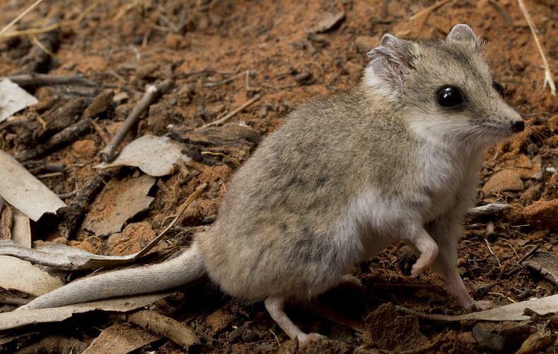
Researchers have successfully extracted and decoded RNA from an extinct animal for the first time. Past researchers have mapped out the thylacine genetic blueprint, in addition to the genomes of other extinct animals; however, these past researches were all focused on DNA, but only RNA can reveal how an organism’s cells actually functioned. The past research mostly focused on DNA because RNA is a relatively fragile molecule, but it is responsible for turning DNA’s genetic instructions into cellular functions and thus must be used to reveal a cell's true biology. The specimen they retrieved the RNA from was a roughly 130 year old Tasmanian tiger, often referred to as a thylacine. Skin and muscle were taken from the desiccated thylacine, then ground into a powder. Chemicals were  then added to isolate nucleotides, the building blocks of RNA. A computer algorithm compared the nucleotide sequences, with a database containing the genomes of thousands of organisms across numerous kingdoms. The results from this research provide new insights on what genes control certain attributes of the organism, for instance, researchers pinpointed RNA molecules that coded cells to make slow-twitch muscle fiber, which helps with endurance. Researchers also found over 250 thylacine-specific short RNA molecules that have sequences that regulate cell functioning. Some scientists  are hopeful that the decoded RNA could aid efforts in bringing this carnivorous marsupial back from extinction. The plan for bringing this species back would involve modifying the genes of one of the thylacine’s closest living relatives, the fat-tailed dunnart. 

I find this research interesting because it can lead to the revival of an extinct species, but after looking at one of the closest living relatives to a carnivore with a mouth that can open over 80 degrees I have my doubts. I find the prospect of learning how extinct animals' genes work interesting, but I feel like this might not be the animal to start with. A species that would be more realistic to bring back would be time better spent in my opinion, but the research had to start somewhere and I suppose that the Tasmanian tiger is just as extinct as any other species that's been wiped out. The thylacine specimen that had the samples taken from it was improperly stored for long term storage. It was just thrown in a cabinet in a bag for over 100 years, so if nothing else this makes me more hopeful that there will be specimens that exist that can lead to the recreation of extinct species.


https://www.sciencenews.org/article/first-time-decode-rna-extinct-animal-tasmanian

https://www.nature.com/articles/s41559-017-0417-y%20

http://m.genome.cshlp.org/content/early/2023/07/18/gr.277663.123.abstract

Tuesday, November 30, 2021

Conservation for Homebody Tiger Sharks


Tiger sharks are undoubtedly one of the most recognizable megafauna that humans come into contact with in the ocean. Researchers recently discovered that tiger sharks have new formed at least two genetically distinct groups in both the Atlantic and Indo-Pacific basins. They found this by comparing the genomes of 242 tiger sharks from various locations around the world. The marine biologists focused on specific genetic markers on their genome and contrasted the differences. One of the authors expected that in the future, the differentiations in their genomes may lead to speciation. 

And while these sharks are generalist predators, their numbers are dwindling from the fin trade and commercial fishing. This is causing them to be listed as a Near Threatened species according to the IUCN. However, studying their genomes may help conservationists develop fishery policies to protect the sharks in each of the areas. Since these "homebody" sharks don't tend to migrate to reproduce as once thought, their habitats in each area can be protected and managed separately. The researchers hope to use these methods to investigate other species of sharks as well.


Monday, November 22, 2021

How Bumble Bees Get Their Iconic Stripes


A recent study published by researchers at Penn State have found the genetic pathways that give bumble bees their signature look. Although many people associate black and yellow with those in the Bombus genus, there are at least 250 species across the world. Across these 250 species, there are an estimated 400 different color patterns that include blacks, whites, oranges, reds, and yellows to warn predators to stay away. Previously, these researchers explained that a major developmental gene, called a Hox gene, activates a "complex set of downstream genes" that drive the pigmentation changes. But they couldn't quite nail down how the change in the Hox gene led to pigment changes.

To figure this out, they looked at which genes were being targeted by the Hox gene. What they found was that by targeting the next major developmental gene, several melanin genes could be altered to reinforce the color traits. Their work also contributed the understanding of genes involved in making the red pigment called pheomelanin. Once thought to only occur in vertebrates (and in humans with red hair!), it was recently found in insects as well. By understanding how these bumble bee pigmentation genes develop, more research can be done on their diversification and evolution.

Thursday, August 5, 2021

The Genetics & Biology in Preeclampsia

The Genetics & Biology in Preeclampsia


    Preeclampsia is a medical condition that occurs during pregnancy, “...characterized by the onset of hypertension, proteinuria, and edema”(Kanasaki and Kalluri). This condition results from placenta defects. It affects approximately 5 to 8% of women during pregnancy. One of the most common and major complications is hypertension (high blood pressure) which is often associated with preeclampsia. This can cause complications to the mother and the unborn child. This condition leads to the presence of edema, which has the plasma volume lower than normal. This may cause damage to the mother and baby’s organs, because of the lower systemic perfusion. Preeclampsia can affect not just the pregnancy, but also the kidneys, because it can cause glomerular endotheliosis. “The risk of preeclampsia increases in those who have limited sperm exposure with the same partner before conception” (Haram et al.).

This condition can cause other medical conditions such as HELLP syndrome, which brings more complications to the pregnancy. In many studies, it shows the “evidence of a genetic association with preeclampsia for the endoplasmatic reticulum aminopeptidases 1 and 2 (ERAP1 and 2) genes. The ERAP1 and ERAP2 genes encode enzymes that play roles in blood pressure regulation via involvement of the renin-angiotensin system in addition to the innate immune system” (Haram et al.). Preeclampsia is still being studied and researched to understand the cause and all of its factors involved.


1. Genetic Aspects of Preeclampsia and the HELLP Syndrome (hindawi.com)

Haram, Kjell, et al. “Genetic Aspects of Preeclampsia and the HELLP Syndrome.” Hindawi. Accessed June 2014.
2. The biology of preeclampsia (sciencedirectassets.com)

Kanasaki, Keizo, and Raghu Kalluri. “The Biology of Preeclampsia.” Biological Science Collection. Accessed August 2009.

Wednesday, March 31, 2021

Fetal Development Powered by Genetics: Artificial Wombs


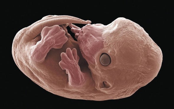
    In the article, “Lab-Grown Mouse Embryos Form Limbs and Organs'' scientists had managed to develop a method that would allow them to grow a mouse embryo outside its mothers uterus. Traditionally when studying the embryonic development of a species; which had allowed scientists to understand how a single cell would develop into a body with specific cell types. To see how fetal development occurs and what important features are present during  different stages surgical images were taken from cutting into the womb of the mouse. This time however scientists were able to artificially create a womb which allowed them to  analyze the gestation of the embryo in real time without having to cut into the womb.

    Having the right conditions to grow the embryo was important to maintain proper functionality and development. Conditions such as the right amount of pressure, oxygen, and nutrients were vital to sustain the embryos genetic material and even managed to get the embryo to develop functioning systems like the heart beating, limb buds, a digestive, auditory and visual system. The lab only managed to keep the mouse alive for 11 out of the 20 day gestation period needed to have a fully developed mouse. This was due to the fact that there still needed to be more advancement in how to supply an artificial bloodline that would more directly aid in the fetus development. Not only has this research produced altering discoveries it has foreshadowed the probability to grow a human embryo outside of the womb as well. Such reasons to do so are important when considering the genetic factors. These genetic factors look at the embryos modes of inheritance and susceptibility of certain conditions based on if it is sex-linked or autosomal dominant or recessive.The reason to even consider growing a human in an artificial womb is to be able to detect any birth defects and diseases that could endanger the developing human embryo. Until then more research will need to be done. 

Thursday, February 11, 2021

New Genetic Blueprint Found in Parasitic Plants


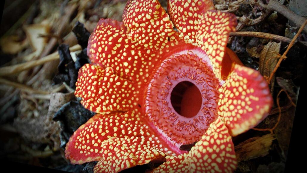
Newfound research on the genetic instruction book of the Sapria genus reveals the lengths to which it has gone to become a specialized parasite. The new discovery illustrates the level of commitment S. Himalayana and its relatives have given to evolving a parasitic lifestyle and provides a comparison to other extreme plant parasites. 

Based on the findings published by Current Biology, most of the Sapria genus have lost half more than half of their genetic material. Not only that but plants like the Sapria Himalayana and their genomes were used for research. Findings showed they had completely removed the need for stems, roots, and even any photosynthetic tissue—and based on further research, even chloroplast genome have vanished

Charles Davis, an evolutionary biologist at Harvard University states that these genetic variations from such parasitic plants have left biologists confused by the sudden change. Such obvious plants recognize by their “rotting flesh” smell, are no longer producing flowers. He notes, “these plants have lost half of their genes, yet they still survive.” 

However, further investigation into this interesting genetic modification will allow researchers to determine some of biology’s limits, which I think will benefit us greatly. 

Links:


Friday, December 11, 2020

Does Genetics Have an Influence on Our Actions?

    In this article posted by Genetic Literacy Project the topic of whether our genetics shape our actions was discussed. It was mentioned that mental-illnesses, vulnerability, longevity, and impulsive tendencies are some-what determined by genes. The ability to analyze a fetus's brain at 20 weeks while still in the womb was mentioned; susceptibilities for conditions such as ADHD, autism, bipolar disorder, depressive disorder, and schizophrenia could be found during this stage. Next, the topic of epigenetics was mentioned, in which the tags connected to genes are analyzed. These tags are placed upon genes and are passed onto offspring. An experiment was mentioned in which two groups of mice were exposed to the smell of cherries; however, in one group the mice were shocked when they smelt the cherries. This resulted in the offspring of the shocked mice being traumatized by the smell of cherries. It was mentioned that there has not been enough research done to relate the same trend occurs in humans. Although it has been found that descendants of US Civil War prisoners are more likely to die younger in life, and Holocaust survivors offsprings having a higher level of cortisol, which is the hormone involved in stress responses. I found this article to be very informative, and I enjoyed the researches that they shared. I agree with the information presented in this study since it was backed up with research. I hope more research is done surrounding the topic of epigenetics. I find epigenetics to be very interesting, as another it connects two of my favorite areas of science: psychology and biology.

Tuesday, October 9, 2018

2018 Nobel Prize Winners for Their Work with Proteins and Enzymes

   
      Last week was a momentous occasion for many people in the field of research, as the Nobel's Prizes for each category were announced. Many people waited and watched, riddled with anticipation, as each winner was announced, and by Monday there were a grand total 12 Nobel Laureates recognized for their outstanding achievements. Among the 12 Nobel prize winners, there were 3 laureates of particular interest that contributed further to the knowledge and understanding of biology and genetic evolution.
     Dr. Frances H. Arnold was the first Nobel laureate to be recognized for her work with directed evolution of enzymes. Dr. Arnold pioneered the first bio-engineering method in 1990 called directed evolution. Directed evolution is a method that introduces random mutations into a gene, which is then inserted into an enzyme and is used as a template to produce a catalyst for desired reactions. Directed evolution has been used to produce chemicals that are safer for people as well as the environment. Chemicals such as bio-fuels, medicine, laundry detergents and more have been created using this process. Dr. Arnold has used evolution to her advantage and in an article in the NY Times she stated "I copied nature’s inventions, this wonderful process of evolution, to breed molecules like you breed cats and dogs.”

       Dr. George P. Smith and Sir Gregory P. Winters shared the 2018 Nobel Prize with Dr. Arnold for their work with bacteriophages. Dr. Smith, from University of Missouri, was looking to identify unknown genes using peptides and bacteriophages. In order to identify the unknown genes, Dr. Smith would insert genes into bacteriophages, which would then express the protein on the surface. Dr. Smith watched these antibodies to see if they would interacted with the proteins and identifying them as a pathogen. The different types of antibodies only fit specific types of proteins, and once an antibody identifies a protein, scientists can then deduce the identity of the unknown gene.
     Sir Gregory P. Winters, from MRC Laboratory of Molecular Biology UK, went a step further and used Dr. Smith's research to produce more efficient and prolific antibodies. Instead of inserting an unknown gene into a bacteriaphage, Dr. Winters inserted the bacteriapages with antibodies and selected the ones that bound the most effectively. He then repeated the process several times to produce powerful disease fighting antibodies. According to the NY Times, "the first antibody drug developed this way, adalimumab, which is sold under the brand name Humira" (Chang, 2018). This drug was used to fight diseases like rheumatoid arthritis, psoriasis and inflammatory bowl disease. In addition to his drug synthesis, Dr. Winter also used this technique to produce efficient antibodies that could shrink tumors in the human body. In order to do this, Dr. Winter tweaked a mouse antibody, using the same technique, and injected that antibody into a patient with a deadly tumor. The tumor shrank successfully and the patient suffered from no side effects. The shrunken tumor was the first ever human test to see if this procedure could be used for the public health.

Friday, October 5, 2018

Breakthrough Discovery Linking Embryos to Tumors.


Developmental biologist Dr. Alberto Rosellió-Díez, from Medicine Institute at Monash University and Dr. Isaac S. Kohane, from MIT-Harvard Division of health Sciense, have been trying to understand how tissue growth, during embryonic development, is related to the cancerous tumors. Studies post in the Scientific American recently linked these two processes together stating that “cancer cells are the same programmed genetic instructions active during various stages of embryonic and fetal development.” Kohane believes that by observing these growth processes, they will be able to have a better understanding of how embryonic development contributes to cancer growth later on in life.

First, to better understand how genetic instructions program tumors to grow in adults, Díez and his team needed to study tissue growth during the embryonic development. In order to do this the team created a model in mice, using prior knowledge of cell modification, to come up with a mechanism that inhibited cell growth. Using this mechanism, the team injected the rear limb of a mouse with a growth inhibitor and watched to see how the rest of the embryo reacted. During this process the rear limb stopped growing and the rest fetus reacted by slowing down its growth as well. It turned out that when the scientists stopped the rear limb from growing, the placenta also reacted by signaling the other limbs to stop growing as well, allowing the slower one to catch up. The placenta, which acts basically as an immune-endocrine organ, mediated the growth process of the other limbs allowing for symmetrical growth of all the limbs. The growth inhibitor allowed the scientists to see for the first time how tissue growth is turned on and off by instructions, programmed during embryonic development. An article in Scientific American discusses these findings in further detail. The results obtained by Díez and his team seem to be promising and they may even help doctors like Kohane explain the effects that these embryonic instructions have on cancer. Kohane, who is currently working with tumors, discovered that gene signatures during the third trimester mirrored growth rates in cancer. Among some of these tumors that exhibited these signatures were ardenocarcinoma, T cell lymphomas, and thyroid cancer. Using these signatures and the research from embryonic development, Kohane hopes to be able to see early warning signs of cancer. Tumors, which are generic terms for neoplasms usually grow rapidly and are able to invade surrounding tissue. Scientists and doctors have observed a remarkable similarity between the behavior of embryos and tumors. Lloyd J. Old, a chairman of the Ludwig Institute for Cancer Research, stated “If you’ve ever seen the trophoblast invading the uterus, it invades, spreads, creates a blood supply. It also suppresses the maternal immune system… All of those are characteristics of cancer.” These new discoveries and observations have given us a much better understanding of embryonic development and its relationship to cancer. However, the research is still in its infancy stage and there is still a lot of speculation on how things work, but with breakthroughs like Dr. Rosellió-Díez and Kohane's research, it might not be long until we can treat cancer much more effectively.  

Monday, March 19, 2018

Molecular cuisine for gut bacteria .


Image result for gut bacteria

Gut bacteria have a very big part in our digestive process as well as our health. Researchers still do not know the kind of food our gut bacteria like to live off of or how they seem to metabolize nutrients. The researchers selected 96 strains from 72 bacterial species, representing the most occurring and abundant species in the human gut. Nassos Typas says in the article, "Our resource provides scientists with tools to experimentally investigate the gt microbiome ecology, going beyond correlations and identifying causes and effects." 

While in the middle of characterising their nutritional preferences for molecules and the gut bacteria the researchers also discovered unknown metabolic features of some other bacteria. Kiran Patil, one of the members of the research team says, "We were surprised to find new bacteria with the capability to utilize mucin, the protein that makes up mucus. These bacteria can contribute to inflammation and infection by weakening the protective mucus barrier lining the gut. Another surprise came from bacteria that proved to be inhibited by amino acids and short-chain fatty acids, common ingredients in most growth media. It turns out that rich media with many nutrients can be toxic for these species, whereas we used to think: the more food, the better." All in all, even closely related bacteria to the gut bacteria sometimes had completely different nutritional preferences.  This new discovery of the gut bacteria will surely impact the medical industry in helping create medicines that are more effective in killing viruses and bacteria in our body that can cause colon cancer or other stomach related issues. 


https://www.sciencedaily.com/releases/2018/03/180319120523.htm
https://www.embl.de/index.php

Wednesday, January 24, 2018

Hermaphroditic roundworms lose half their genome from sister species when self fertilizing


An article done by the New York Times describes how tiny roundworms, Caenorhabditis briggsae and Caenorhabditis nigoni are extremely similar genetically, but very different in their sexual reproduction processes. In order for C. nigoni to mate, there needs to be a male and a female for fertilization to take place. This is different for C. briggsae however, in that they are able to self-fertilize by producing sperm to fertilize their own eggs. Because C. briggsae are hermaphroditic, there is no need for sexual reproduction. This genetic adaptation has occurred because there is a lack of males in the population. In a study published in Science in the beginning of January of 2018, biologists found that since the hermaphroditism took place in the one set of roundworms, it lost half of the genes since it broke away from C. nigoni approximately a million years ago. A study done by the University of Maryland-College Park concludes that this loss of genes is related to the male population which is dwindling in numbers. When introduced to the male secreted short genes (m.s.s.), C. briggsae produced more offspring than the hermaphroditic organisms. Dr. Haag from the University of Maryland-College Park suggests that these organisms leaning towards hermaphroditic lifestyles may “put a break on population”.


This article I found very interesting. It’s neat to see how hermaphroditic traits in one roundworm compared to their ancestral sister alter their entire genome. Although the roundworm C. briggsae do not yield as many offspring through self-fertilization unlike if they were to be in contact with a male, it provides evolutionary traits in that population growth and size can be halted/reduced. It’s also hard for these worms to find mates as well, which make it hard for new colonies of C. briggsae to get started.

(Related Article)

Friday, April 15, 2016

Scientifically Built Bacteria?

Craig Venter and scientists have recently constructed a bacteria, which has a genome consisting of 473 genes.  This appears to be the smallest number of genes necessary for a viable organism.  Scientists used Mycoplasma, which was known to be one of the most genetically simple, to build the bacteria.  In nature, Mycoplasma myocoides contains 901 genes.  So, compared to the 4,000 - 5,000 genes used in E. coli, 473 is a very small number.  Researchers replicated the Mycoplasma genome and inserted it into a Mycoplasma capricolum.  The organism was named syn3.0.  Scientists hope to study syn3.0 in order to discover more about general biology, and they hope to use the bacteria for drug and chemical production.
Venter and his colleagues initially expected to be able to use as few as 300 genes to create a completely viable bacterium.  However, the bacteria required 473, and this has led to a number of questions regarding the function of the genes.  Scientists are currently unable to determine the purpose of about 1/3 of the genes in syn3.0.  All they know, for sure, is that the genes are essential to life.  Venter and his fellow scientists believe that 41% of unclassified the genes contribute to genome expression, 18% code for cell membrane structure and function, and 7% is responsible for preserving genomic information.  

Constructing man-made life is always going to be a controversial topic.  In a world that where religion is still vital to a large number of people, there will be arguments about the ethics of this field of science.  Questions like, "who are we to create life?" will surely be asked.  However, the scientific findings could be humongous.  If we are able to manipulate the genome easily, we could possibly create cheaper more effective pharmaceutical drugs.  Further, we can learn more about the genes that are necessary for syn3.0 to survive, and we can understand similar gene's significance in other bacterial cells.  

Thursday, April 14, 2016

Potential Alzheimer's Disease Treatments Using Suppressor Genes

Scientists from The Scripts Research Institute have identified a suppressor gene in Drosophila brains. According to these scientists, drosophila are a recognized substitute for human brain and memory studies.  One researcher, Ron Davis, collected and screened about 3,500 fruit fly genes, and they observed several memory suppressor genes that the brain uses to filter information.  The gene also allows the brain to differentiate important memories from less important ones.  When a specific gene was identified (DmSLC22A), it was removed from the drosophila genome.  The researchers noted that flies' memory capability nearly doubled.  Davis stated, "The fact that the gene is active in the same pathway as several cognitive enhancers currently used for the treatment of Alzheimer's disease suggests it could be a potential new therapeutic target." So, Davis asserts that the gene could help enhance memory of Alzheimer's patients.  It may seem that drosophila and humans are too evolutionarily different from one another, but according to Davis, memory processes are still relatively similar between mammals and fruit flies.
Alzheimer's disease is a fatal disease that has a genetic component.  Early Onset Alzheimer's Disease can be caused by single-gene mutations on Chromosomes 1, 14, and 21.  When the mutations manifest themselves, they cause abnormal proteins to be created.  When the genes are working normally, they work together to create amyloid precursor protein.  However, when the mutated genes are turned on, amyloid precursor protein cannot be made properly.  The variations of amyloid precursor genes can be detrimental to brain health, and they are characteristic of Alzheimer's disease.

Advances in Alzheimer research are a great achievement in medicine.  However, I am somewhat skeptical of the validity of the Davis study.  While Davis states that humans and drosophila have similar memory processes, I think that claim may be too bold.  Humans and drosophila are extremely far apart evolutionarily, and I find it hard to believe that our brains function that similarly.  However, any findings in Alzheimer's are welcomed, and they should be explored further.  If the research proves to be accurate, this could be a major step forward in preventing memory loss in patients afflicted with the disease.

Thursday, January 29, 2015

Complex Life's First Milestone

Hydrothermal vent at the bottom of the ocean
     In regards to the origins of complex life forms, one theory is globally accepted. This is the theory that RNA strands were being continuously formed in the mixing pot that was the prehistoric ocean. These strands would grow and reproduce until longer, more complex chains formed actual life-forms such as early amoebas and worms. Scientists have now discovered how it may have been possible that complex RNA strands out-competed simple, shorter ones.
     Technically speaking, shorter RNA strands can reproduce more efficiently because, although they are more primitive, with less material to copy the process is quicker. However, an article in Science Magazine claims that pores in the rocks surrounding hydrothermal vents in the oceans floor may have created the perfect breeding ground for the complex RNA strands, that eventually lead to us. The superheated water that emerges from these fissures is highly enriched with nutrients. Through a physical effect known as thermophoresis, “charged molecules in a solution accumulate in colder water, and the longer chains, having more charge, would do this more often than shorter chains”. Add this save haven, porous environment, together with the flow of nutrients and temperature changes from hydrothermal vents, and the result is much more efficient reproduction.
     This theory was recreated in a laboratory experiment that resulted in the longer strands reproducing much better inside the pores, leading to larger and more stable populations. The new findings may have opened a door to better understanding the evolution and origins of complex life, and the environments that made it possible.  

Monday, April 21, 2014

Why alcoholism saps muscle growth


In this article, mitochondria fusion in muscle cells was discovered. The researcher team led by Director of Jeffersons MitoCare Center showed that the mitofusin fusion proteins, was most important in skeletal muscle cells. They tested whether mitochondrial fusion was the culprit in examples of muscle weakness, such as alcoholism. The research showed that the abundance of Mfn1 went down as much as 50 percent in rats on a regular alcohol diet, while other fusion proteins were unchanged. This decrease was coupled with a massive decrease in mitochondrial fusion. Mfn1 and mitochondrial fusion was linked to increase muscle fatigue. Alcohol can have a specific affect on this one gene involved in mitochondrial fusion suggest that other environmental factors may also alter mitochondrial fusion.

I believe that alcohol plays a huge impact on the way people develop muscle as well as fatigue in different exercises.  I have experience in both of these actions and going to the gym the day after consuming alcohol, my body feels terrible. I lose my breath much faster and cannot produce as much as I would on a normal day.


Gut Microbes Gobble Cocoa


In this article, Microbes like bifidobacterium and lactic acid bacteria that reside in the human gut feast on chocolate. It was said that when you eat dark chocolate, these bacteria grow and ferment it, producing compounds that are anti inflammatory. In the human digestive tract, food scientists tested three cocoa powders. Researchers found that within the mock gut, cocoa is fermented and the large polyphenolic polymers are metabolized to smaller molecules. Gut microbes could help reduce a persons feeling of hunger after digesting cocoa. The fiber is broken down into short fatty chain acids, which are absorbed and could have an affect on satiety. Researchers have yet to confirm results meaning do not go to your local candy store and start eating all the chocolate bars in sight, many of these products contain sugar and fat which is not good for you.

http://www.landesbioscience.com/journals/gutmicrobes/

Saturday, April 19, 2014

Plants evolve ways to control embryo growth

In this article, plants have evolved ways to control embryo growth and development by emitting information from surrounding cells. Female sex cells and placenta like endosperm contained within plant seeds send out specific signals to developing embrtos to help with there growth. This new information changes changes are understanding of plant development whci could allow for better breeding of plant types. Because of the changes in the environment as well as our climate, there is always need for more plants in the aid of oxygen as well as many other areas. Plantembryos are found within seeds and once germinated, they give rise to an adult plant. Before the discovery, the ability of non emryonic plant cells to direct embryo groth was unrecognized, but now they have valuble information that neighoring cells can directly act with and influence embryo cells. The next step is to indentify the embryonic factors that respond to thses non embryonic signals and understrand there mode of action.

http://phys.org/news/2014-04-free-seed-ossi-nurtures-patent.html

Some immune cells only defend one organ

In this article, scientists have discovered a new way the immune system may fight cancers and viral infections. Research has been done on mice and in mice it shows that some organs have the immunological equivalent of "neighorhood police" speacialized squads of defenders that patrol only one area, a single organ, instead of an entire body. If one group of cells absolutly needs a transcription factor to exist, while another group of cells dosent care if that factor is gone, that strongly suggests that the two groups of cells use disticnt developmental pathways and are therfore different. The results point to at least four types of natural killer cells rather than just one major type long recongnized by immunologists. After discovering this new addition to the body this changes some of the concepts and thoughts of many reaserchers and may help with different cancers.


http://www.nature.com/icb/index.html

Friday, April 18, 2014

Breaking Bad Mitichondria

In this article, researchers have identified a mechanism that explains why people with the Hepatitis c virus get liver disease and why the virus is able to persist i the body for so long. The pathogen attacks the liver cells energy centers, mitochondria, which does not allow for the cell to fight off infections or diseases. Hepatitis c causes liver cancer. After the mitochondria are done being attacked the proteins tell the mitochondria to eliminate the damaged area, but the process to repair ends up aiding the virus making things worse. Mitochondria convert food onto a form of energy which is used by the cells. The virus stimulates the production of protein that induces viral damaged mitochondria. Although mitochondria help with fighting off the infections and diseases it also helps with keeping the virus infected cell alive. The virus is able to use the mitochondria to keep reproducing new cells and amino acids to help fuel its continuous replication and virulence. Understanding mitochondria is what keeps the virus going when attaining the hepatitis c disease.


http://jcs.biologists.org/content/123/9/1389.abstract