Showing posts with label "Neurons". Show all posts

Sunday, December 8, 2024

Can Kidney Cells Make Memories?


Not quite. Kidney’s can make memories on a molecular level but not the type of memories we commonly think of. Kidney cells, like neurons, can store information and recognize patterns. The researcher, Nikolay Kukushkin, says that this does not contradict current knowledge on memories, it simply adds to current knowledge because they do not act like neuron memories. Other cells, like neurons, need to keep track of stuff that is going on which they do using a protein central to memory process called CREB. For a little background in memory in neurons, a chemical signal passed through them which then begins CREB production. CREB activated more genes which change the cell and start the molecular memory machine.

Through their experiment of inserting embryonic kidney cells with a glowing gene found in fireflies, they activated memory instructions in the kidney cell. The kidney cell responded to artificial chemical pulses which are like the signals which trigger memory in neurons. Different lengths of pulses triggered various levels of responses, or lighter. It is believed the newfound idea that cells can be given more complicated memory tasks, that this has a potential to introduce new potential drugs to treat disease, specifically memory loss related.

This has enormous potential, and I would love to see where it goes. It is currently just a thought and a small step in proving potential of this finding. I do not see why other cells could not mimic the process of neurons because at a simple level neurons are just specialized cells, so it makes sense that these kidney cells could perform this. It would be interesting to see this application in medicine or aiding those with memory loss.

Links

https://www.sciencenews.org/article/brain-kidney-cells-memory

https://www.hopkinsmedicine.org/health/wellness-and-prevention/inside-the-science-of-memory


Monday, November 13, 2023

Study Connects Neuron Gene Expressions to Differing Functional Distinctions

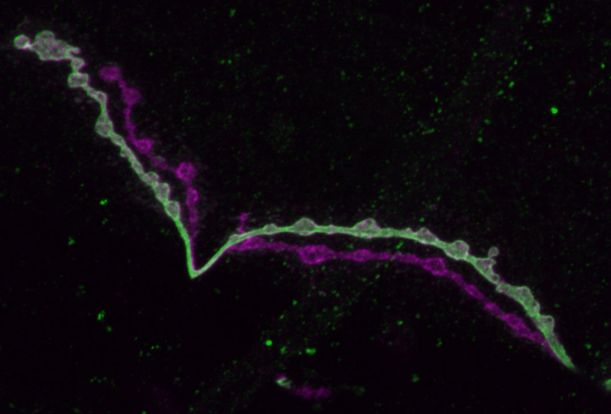
 Long, thin, bumpy extensions of two neurons extend from upper left to bottom right across a black background. One is light green, the other is magenta. The light green one features patches of darker green amid little bumps along its length.

In a new study in Neuron, neurobiologists at the Picower Institute for Learning and Memory found that two closely related neuron subtypes in drosophila differed from each other in how they expressed more than 800 genes, about 5% of the total amount of genes in the fly genome. The two neuron types studied were both from what could be considered the spinal cord of the fruit fly. These neuron types control the muscles by releasing the neurotransmitter, glutamate. The main functional differences of the two subtypes are that the “phasic” neurons connect to a lot of muscles and emit big, occasional bursts of glutamate, and the “tonic” neurons connect to only one muscle and emit a constant, small amount of glutamate. Phasic neurons make fewer synapses on an individual muscle than tonic ones do, but make about 4 times as many synapses in total because they innervate so many more muscles. Tonic neurons have more inputs from other neurons due to it having more widely branching dendrites. Of the expressed genes of the neuron types, a significant amount helped with the growth of the axon branches, some helped with the structure and function of synapses, and others played a role in the types of chemicals the neurons were sensitive to as inputs. Researchers disrupted the functions of some genes to see which were the most different between the two subtypes. By disrupting the Wnt4 gene, a gene expressed 40 times more by the tonic neurons, the synaptic growth decreased significantly in the tonic neurons. By mutating a calcium ion buffering gene found 30 times more in phasic neurons, the phasic neurons had higher resting calcium levels similar to tonic neurons.

This study holds a lot of significance in not only discovering how different genes can overlap and differ in different cell types, but also specifically in how neuron subtypes can differ. The research presented in this article is exciting because figuring out how different kinds of neurons develop from their expression of different genes helps in advancing how a brain works. It could also help in understanding what can change or go wrong in disease. The study compares two similar cells in a very detailed manner and shows that even similar cells can have a lot of differences in gene expression to develop specific, distinct functions. 

https://news.mit.edu/2023/study-connects-neural-gene-expression-differences-functional-distinctions-0825  

https://news.mit.edu/2020/two-seemingly-similar-neurons-show-distinct-styles-in-muscle-interactions-0720#:~:text=The%20%E2%80%9Ctonic%E2%80%9D%20neuron%2C%20which,spring%20the%20muscles%20into%20action

Sunday, October 22, 2023

Genetic Basis of Postpartum Depression

PPD, GABA, and the Thalami


    A recent study from the UNC School of Medicine revealed that 14% of the variability in postpartum depression (PPD) is due to genetic factors/on a genetic basis and also provided a glimpse into the connections PPD has with a variety of pre-existing psychiatric disorders. The basis of PPD symptoms lays with GABAergic neurons in the hypothalamus and thalamus; areas of your brain responsible for regulatory functions (hunger, thirst, thyroid function, etc), and seem to have promising advancements for future PPD research. This data was collected using a genome-wide-association study and compiled data in order to get a bigger glimpse into PPD. Jerry Guintivano, PhD, assistant professor of psychiatry at the UNC School of Medicine, was able to identify similarities in the genetic architecture of PPD and major depression, bipolar disorder, anxiety disorders, post-traumatic stress disorder, insomnia, and polycystic ovary syndrome, meaning "...PPD symptoms likely occur as a result of the interplay between the same genes involved in these other psychiatric and hormone-related conditions." Furthermore, the research concluded a link between PPD, its genetic architectural siblings, and GABAergic neurons in the hypothalamus and thalamus of the brain: neurons releasing GABA neurotransmitters appear to have a link to PPD and its similar familial connections of disorders. Brexanolone is a common drug used to treat PPD and now researchers are able to see the interconnectedness of Brexanolone and the two regions of the brain, which is promising for additional treatment in the foreseeable future. Due to such a wide study and too few PPD cases, however, the researchers at UNC were unable to identify specific locations on the genome or loci that are consistently similar amongst the data group.

    This study was fascinating to read and it provided an overlap between multiple specialties in biology -- from neuroscience to anatomy to physiology. Women's health is making big strides in the 21st century and I think this article is just another step towards more progress being made. The linkage between hormones and psychiatric disorders seems to be a hot topic, and scientists had avoided looking at female models for a long time due to the "complexity" of female hormones and their interactions with physiological processes in the body. But now, we have more evidence that encourage this research and should make women feel a lot better about the improvements being made and the connections established between the two factors. Furthermore, the researchers had also confirmed that PPD is genetic which is also a huge step as well! This can impact treatment plans for pregnant women and might improve their quality of postpartum treatment if they are better prepared and more aware of their chances for PPD. Nevertheless, this study was so interesting to read about and I love the idea of its medical/pharmaceutical improvements as well. 


LINKS:

1) https://neurosciencenews.com/post-partum-depression-genetics-24968/ 

2) https://news.unchealthcare.org/2023/10/researchers-confirm-postpartum-depression-heritability-home-in-on-treatment-mechanism/ 


Monday, September 19, 2022

A mutation that may have given Homo sapiens a neurological advantage over Neanderthals


 A group of molecular biologists and geneticists in Germany had an interesting breakthrough when studying differences between the brains of Homo sapiens and Neanderthals.  Although scientists have known for a while how the brain sizes of these two hominin species compared, there was not much evidence about the structure or neurology of the brain. Now, however, these German scientist have identified a key genetic mutation between the two. Modern humans have a mutated version of the TKTL1 gene found in Neanderthals and it appears to allow for a more efficient creation of neurons in the brain. A great number of neurons could have contributed to an increase in neuroactivity/intelligence and therefore Homo sapiens' evolutionary success as opposed to Neanderthal's extinction. That would be incredibly significant for a mutation that is only different by one amino acid.

Other scientists are a little more hesitant to accept this proposal. The article discusses how this researched focused on only one cell line and that they largely ignored data that was not Western European. These critics are not turning down the proposal altogether. They are simply calling for wider and more extensive studies that could contribute to this finding.

Personally I think the concept of this research is very fascinating. One of those big life questions that I have is why out of all the early hominin species, we were the only ones to make it this far. This article exposed me to a potential contributing factor and gave me hope that more could come soon. Overall, I think the article was well written. I do wish, however, they had talked more about how the German scientists accessed the Neanderthal DNA to compared it to modern humans.


Tuesday, March 12, 2019

Sleep Tight! Researchers identify the beneficial role of sleep

Image of chromosome dynamics(red) and neuron activity(green) in Zebrafish
Research has been done and published by the Journal of Nature Communication which studied the pattern of sleep disturbances and sleep affect our brain performance, aging, and it's roles in brain disorders. Researchers were able to successfully isolate and identify sleep in an individual chromosome using 3D time-lapse techniques. These images show how single neurons need sleep so that they are able to perform nuclear maintenance and while we sleep DNA damage levels are normalized because, while we are awake DNA damage consistently accumulates throughout the day and repairing it is less efficient while we are awake.

I think it's interesting to see how something as normal as sleep plays an important role in not only how we function on a day to day basis but, in how our brain develops. It's funny because usually, you hear people saying how without sleep they are not themselves and it's literally true.

Tuesday, December 11, 2018

Regrowing damaged nerves hinges on shutting down key genes


Quadriplegia is the partial or complete paralysis of both the arms and legs that is usually due to injury or disease of the spinal cord in the region of the neck.

Neurons in the brain and spinal cord don't grow back after injury, unlike those in the rest of the body. Now, researchers have identified some of the key steps taken by nerves in the legs as they regenerate. The findings lay out a path that spinal cord neurons might be able to follow, potentially leading to improved recovery for people paralyzed by spinal cord injuries.

Researchers at Washington University School of Medicine in St. Louis have been working with mice and have identified some of the key steps taken by peripheral nerves, which are those in the arms and legs, as they regenerate. There's no way to reverse a spinal cord injury that has already occurred. The neurons that form the spinal cord do not spontaneously heal themselves.

they have found a set of genes related to sending and receiving chemical and electrical signals, the primary duty of mature neurons , this had to be silenced for the injury to heal, the researchers showed.

The idea that cells must become less mature in order to regenerate is not new, but Cavalli and Oh's study provides evidence in support of that idea. The researchers identified the key molecular and genetic players involved in regressing to a less mature state, and showed that the timing of the regression was crucial to successful recovery.

The scientist are still trying to develop a more detailed understanding of when and for how long specific genes must be shut off.


Source:

https://www.sciencedaily.com/releases/2018/12/181210150618.htm Science Daily

https://www.merriam-webster.com/medical/quadriplegia

Monday, November 19, 2018

Improved Diagnostic Techniques for Autism

Autism spectrum disorder (ASD) is characterized by communication and behavior development deficiencies in comparison to peers. These symptoms exist in a wide range and are typically used in diagnostic tests. The earlier patients are diagnosed and treated according to their specific set of symptoms, the better the odds are for healthy social development. Apart from observing behavior, there has not yet been a way to diagnose patients, which can delay treatment start time in some cases. Scientists at Kanazawa University have been working on a technique to utilize brainwave activity to identify ASD symptoms.

The first step to developing this new technique was expanding on their formed hypothesis that autism results from an imbalance in excitatory and inhibitory neuron ratio. A neurotypical individual would posses a ratio of 4 excitatory neurons for every inhibitory neuron.This ratio can be visualized in patients by observing a specific type of brain wave activity referred to as gamma oscillations. Researches studied gamma oscillations in groups of children with and without ASD from ages 5 to 7. Subjects performed specific tasks while being evaluated in response time and brain wave activity.

Scientists expected reaction time to be visibly reduced, but the interesting find came when observing the gamma oscillations. Specifically, a lower frequencies in these brain waves were found, which correlates with reduced inhibitory neuron activity. This finding is characteristic of ASD, and manifests as patients inability to control motor movements at the level of their peers. Observing gamma oscillations, along with current behavioral tests, will assist future patients in earlier diagnosis and therefore better treatment options.

Sunday, April 8, 2018

Genetics and Synesthesia


People typically describe the world around them using the five senses: touch, taste, hearing, sight, and smell. The divisions between the senses become blurred with synesthesia, a condition in which sound (or another sense) may invoke color or flavors or any other typically not linked reaction. A team of scientists have recently been looking into a genetic component behind synesthesia, as it seems to run in families. However the condition is rare in the population making it hard to pinpoint and each person with synesthesia reports slightly different experiences. Using a technique called whole-exome sequencing, protein producing genes of three families with synesthesia were examined against members who were unaffected. As of the time of the article "37 genes of interest" have been marked for further study, six of them dealing with how neurons connect in the brain.
Synesthesia is a complex and fascinating crossing of the senses and has such variability that every person who experiences it has a different account. Discovering how this happens in the brain, and why this happens, would be delving deep into how we process the world around us. The research may also be linked to other sensory related conditions.

Article Link- https://www.smithsonianmag.com/smart-news/researchers-begin-unravel-how-some-people-see-sound-and-hear-color-180968374/
Scientific Paper- http://www.pnas.org/content/early/2018/02/27/1715492115

Sunday, March 18, 2018

Stem Cell Therapy


 The Potential of stem cells in the treatment of traumatic brain injuries

Image result for TBI

Traumatic brain injury (TBI) is a global public health concern, with limited treatment options available. In the U.S alone, between 3.2 -5.3 million people suffer long-term cognitive impairment as a result of TBI. When an individual has sustained a heavy blow to the head it could lead to long-term deficits involving sensory-motor and memory functions. However, the brain harbors neural stem cells that it uses to self-repair itself after damages have been sustained. Unfortunately, these neural stem cells are limited and if the impact to the head was strong it would result in a chronic injury. Therefore the brain would not be able to have a full recovery which would result in future health problems for an individual. As of today, many scientists have begun working with stem cells in hopes of finding a treatment for TBI.
Embryonic and Induced Pluripotent stem cells have acquired a lot of population due to there plasticity and ability to differentiate into any lineage in the nervous central system. Embryonic stem cells (ES) are obtained from fetal or embryonic brains and are strongly considered for neural transplantation because when implanted into a recipients brain these cells can differentiate, migrate, and make innervation to aid the damaged brain to recover. Induced pluripotent stem cells (iPSCs) are obtained from patients themselves and have the potential for autologous transplantation and avoiding ethical and graft rejection concerns. Induced pluripotent stem cells have allowed scientists to explore manipulating this highly plastic population. These somatic cell-derived iPSCs can provide large quantities of pluripotent cells that have high plasticity generating cells for all three germ layers including neurons and glial cells.
These unique properties of Embryonic and Induced pluripotent stem cells have raised hope that many neurological diseases including TBI might be cured or treated.

  Image result for embryonic stem cells

https://www.ncbi.nlm.nih.gov/pubmed/29372464

https://jamanetwork.com/journals/jamaneurology/fullarticle/795390