Showing posts with label embryonic stem cells. Show all posts

Sunday, July 23, 2023

Human Embryo Models From Stem Cells


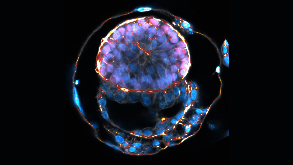
This picture of an embryo model from Jacob Hanna's lab in Israel is one of the few closest lab-grown embryo models to ever exist. These models come from stem cells and are the closest looking cells to early embryonic development because of their possible yolk sac, formation and shapes of cells, and possible placental-like cells. With this advancing technology, scientists begin to question how far they could grow an embryo in a lab. Depending on your country, there are regulations to determine if a scientist could even attempt to achieve a lab-grown human embryo. 

There is a lot of controversy on whether embryo-like structures are useful for real research or are just being used like a game of how far one can one grow the model. Embryo models represent post-implantation stages of embryonic development. During this very early stage of pregnancy is when many pregnancies fail. Researching a human embryo model could teach scientists a lot about why these failures and developmental disorders happen and guide researchers in how to fix them. Although, if this technology gets into the wrong hands, children could be born without a sperm or egg which groups with opinions on cloning. 

Thursday, May 2, 2019

Embryo cells from skin cells

An article at ScienceDaily summarizes the findings of a research conducted at the Hebrew University of Jerusalem (HU). With this study, researchers have found a way of transforming skin cells into the three major stem cell types that comprise early-stage embryos, (embryo, placenta and extra-embryonic tissue), using mouse cells. This was achieved by using retroviruses to insert a series of genes into the skin cells.
In a different article, the author notes that human skin cells have already been tested, and were successfully transformed using the same techniques used in mouse cells. Although successful, the technique with retroviruses could cause tumors in the tissue. The next step is finding a way of altering or switching genes existing in the cells, instead of inserting new copies.

Scientists say that in the future, it could be possible to create whole human embryos out of skin cells. The findings are significant since embryonic cells can be used to model and study embryonic diseases and placental dysfunctions as well as other complications without using "real embryos" or oocytes; therefore escaping some of the ethical debates that surround the field. With this I think that humans are getting closer to find a way of creating life without even the need of sex cells.  

4-cell stage mouse embryos

Sunday, November 20, 2016

A New Technique Allows Insertion of Genes in Non-dividing Cells

Until recently, the only method for modifying DNA was the CRISPR-Cas9 system. However, the new Salk technology is much more efficient than any other method because it allows the incorporation of DNA into non-dividing cells. The cells of the heart, brain, eye, and pancreas do not divide again and are locked in G0. In order for this to be accomplished a specific pathway called NHEJ was targeted by researchers. This pathway repairs routine breaks in DNA sequences by rejoining original strand ends. Researchers constructed a new complex called HITI, homology-independent targeted integration, used to insert genes into the cells. The complex is made up of different nucleic acids. Then, they used a virus to implant the HITI inside the cell to transfer the genetic information to neurons that were derived from embryonic stem cells from humans.  Scientists then completed a genetic modification in adult mice and it was successful. After that, they tried to use the technique as a gene-replacement therapy method. The researchers decided to work with mice that were born blind. They successfully delivered the gene Mertk, one of the genes that, when damaged in humans, causes vision loss. The gene was inserted into the genome of the mice at just three weeks old and by the time they were eight weeks old the mice showed response to light. Furthermore, testing was continued and healing was observed in the retinal cells of the mice. I feel that many people will benefit from this new gene-replacement therapy process. Individuals can be assisted by a gene when having a disability, such as severe eye loss. Also, one that suffers from serious head trauma can gain some of his or her brain cells back. Consequently, he or she will recover from a concussion quicker. This is only the beginning of the great things science can do to help people suffering from genetic damage. Gene therapy is going to revolutionize healthcare in the future. 


Tuesday, March 29, 2016

Behind Each Breath, an Underappreciated Muscle

In order to breathe in, we must flatten the dome-shaped diaphragm; to breath out, we let it relax again. The diaphragm delivers oxygen to us a dozen times or more each minute, a half-billion times during an 80-year life.        
Before the evolution of a diaphragm, our reptilelike ancestors probably breathed the way many reptiles do today. They used a jacket of muscles to squeeze the rib cage.

Scientists suspect that the diaphragm evolved through some change in the way mammal embryos develop: Mutations caused certain embryonic cells to grow into an entirely new muscle. Dr. Kardon and other researchers are trying to understand that shift and why the muscle sometimes fails to develop, with catastrophic consequences.
Original Article:http://www.nytimes.com/2015/04/07/science/behind-each-breath-an-underappreciated-muscle-the-diaphragm.html?rref=collection%2Fsectioncollection%2Fscience&action=click&contentCollection=science&region=stream&module=stream_unit&version=search&contentPlacement=10&pgtype=sectionfront

Sunday, March 22, 2015

Rabies Resistance

   A new technology to determine the resistance or sensitivity to the rabies virus has been developed. Researchers at Texas A&M AgriLife were able to create a platform so that they could look further into how pathogens interact with human genes which will allow them to find genes that are either more sensitive or less sensitive to the rabies virus. The rabies virus is still a concern in today's world, it is the cause to nearly 55,000 deaths per year worldwide. Further understanding the way that genes are affected by this virus will greatly help the human race when it comes to the Rabies virus.
   
     These researchers used "knockout mice" which are laboratory mice that have inactivated existing genes, these genes are replaced with an artificial piece of DNA. They took thousands of different stem cell lines carrying different mutations and separate them into neurons. With this they were able to see that genes that are known to be involved in rabies when mutated had different sensitivities to the rabies virus.
      The experiment was set up so that about 100 embryonic stem cells were spread out on different plates, separating them into fully functioning neurons. The rabies virus was then added showing up as a florescent green color and monitored for several days. The results were astounding, some plates showed large amount of fluorescent green, while others showed very little. The plates that showed high amounts of green were more sensitive to the rabies virus, the ones showing little green were more resistant. The researchers were able to uncover 63 different host genes that will help with new pathways that can aide in the treatment of rabies. This is a remarkable discovery as it will help with other diseases such as tuberculosis, botulism, Ebola as well as many other disorders. This experiment can be used  to help find resistances or sensitivities to almost any virus or bacteria.

Article: Stem cells help researchers peg rabies resistance
Article 2: Rabies

Sunday, November 9, 2014

Neurons Derived from Human Stem Cells Reduce Seizures in Mice

Stem cell derived interneuons integrated into an epileptic mice's brain. 

Scientists at McLean Hospital and Harvard Stem Cell Institute have developed a new strategic approach in helping patients who suffer from epileptic seizures. Associate Neurobiologist Dr. Sangmi Chung successfully transplanted human neurons derived from embryonic stem cells into the brains of mice with epilepsy. The neurons had a special seizure-inhibiting factor. Once the neurons were transplanted into the mice, "The transplanted neurons begin to receive excitatory input from host neurons and in turn generate inhibitory responses that reverse the electrical hyperactivity that cause seizures;" explained Dr. Chung. Half of the mice in the study did not show any signs of seizure activity. The other half of the mice studied showed a diminished amount of seizure frequency.

Before testing on humans, further studies on primate must be conducted. However, the research done so far is very promising. Embryonic stem cells can be differentiated into many other cell types, even when they become neurons. For this reason, the neurons must first need to be purified before entering a human’s brain to ensure that only the interneurons (seizure-inhibiting) are transplanted. This is simply as a precaution to guarantee that cells transplanted into humans are safe without any risk of causing harm to the patient. The researchers are currently working on a method to extract only interneurons from their derived stem-cell neurons.

Approximately 65 million people suffer from epilepsy. Of those, many receive anti-seizure medication. However, approximately 33% of epileptic patients cannot reap the benefits from anti-seizure medication. The medication fails to decrease the seizure activity in their brain. Those who can’t take medication have the option of having the seizure-causing area of their brain removed. It is believed that people with epilepsy have minuscule amounts of interneurons in their brains. This new research takes a step forward for those patients who can’t take anti-seizure medication. It is definitely a new therapy that hopefully makes progress in the years to come.


Tuesday, October 14, 2014

Embryonic Stem Cell Therapy Reports of Long-Term Safety to Treat Human Disease

The Lancet recently published new research in which the first evidence of "medium-term to long-term safety and tolerability of transplanting human embryonic stem cells (hESCs) in humans has been revealed by scientists." The study was conducted on 18 patients who had severe vision loss. They received hESCs and appear to be safe 3 years post-transplant. More than half of the patients experienced restoration of some sight.


"Embryonic stem cells have the potential to become any cell type in the body, but transplantation has been complicated by problems including the risk of teratoma formation and immune rejection," Professor Robert Lanza, Chief Scientific officer at Advanced Cell Technology in the USA. "As a result, immunoprivileged sites (that do not produce a strong immune response) such as the eye have become the first parts of the human body to benefit from this technology."

The participants had one of two different types of eye disorders, half having Stargardt's macular dystrophy and the other half having dry atrophic age-related macular degeneration. Both of these conditions have no effective treatment and they can both lead to complete blindness. The hESCs were differentiated into retinal pigment epithelium cells and were injected in different doses. Some patients received 50,000 retinal cells, some 100,000, and some 150,000 cells. These cells were placed into the space under the retina of the eye (the area with the worse vision).


The hESC cells were accepted and tolerated for "up to 37 months after transplantation." If the patients experienced any adverse effects, after close analysis, it is safe to say that they were not caused by the hESCs. Additionally, 10 of the 18 patients claim to have had significant improvement in their vision in the eyes that received the stem cell treatment.

"Our results suggest the safety and promise of hESCs to alter progressive vision loss in people with degenerative diseases and mark an exciting step towards using [these] stem cells as a safe source of cells for the treatment of various medical disorders requiring tissue repair or replacement," co-lead author Dr. Steven Schwartz, Jules Stein Eye Institute.

Dr. Anthony Atala, director of the Wake Forest Institute for Regenerative Medicine, states how this study is a "major accomplishment" but also that "much work remains to be done before hESC and induced pluripotent stem cell therapies go beyond regulatory trials, but the path is now set in motion."

I absolutely loved this article because I have always had great interest in stem cell therapy. This is a huge step forward for the practice and potential use of hESCs as common treatments for certain disorders or anything else requiring the repair or replacement of tissues. I'm very excited for what is upcoming in the medical field because of advancements such as this in the area of stem cell research and therapy.

Article: http://www.sciencedaily.com/releases/2014/10/141014211709.htm 
Related Article: http://health.usnews.com/health-news/articles/2014/10/14/embryonic-stem-cell-therapy-shows-long-term-effectiveness-safety 


Neuroscientists Make Breakthrough for Studying Alzheimer's

Researchers at Massachusetts General Hospital in Boston have made a breakthrough in studying Alzheimer's. What they call "Alzheimer's in a Dish," neuroscientists Doo Yeo Kim and Rudolph E. Tanzi have replicated human brain cells in a petri dish that mimics the network of neurons in a person with Alzheimer's.

Neuroscientist Doo Yeo Kim(Left) and Rudolph E. Tanzi(Right)
The findings started off as a mere suggestion by Doo Yeo Kim to his colleague Rudolph E. Tanzi. The suggestion was to grow human brain cells in a gel. From there, the scientists added genes for Alzheimer's to the neurons. After a few weeks of incubation, the researchers noticed the development of plaque and tangles, which are key features of the neuron network in a person with Alzheimer's. The growth of the neurons began with human embryonic stem cells. Growing them in the presence of certain chemicals allowed the stem cells to turn into neurons. The only requirement then after was to introduce the Alzheimer gene beta amyloid.

Plaque - pictured an orange,  disrupting a network of neurons - pictured in green, in a petri dish. 
Scientists from all over the country are recognizing the breakthrough Dr. Kim and Dr. Tanzi have made. This is a big step forward for the study of Alzheimer's. This allows an accelerate process of drug testing for the disease. Before scientists had to rely on mice to test newly synthesized drug treatment which took a minimum of a year to study. However, with the petri dish method, researchers can study the effects of drugs within a matter of months. The only downfall of this discovery is that the replica does not take into consideration immune system cells, which are known to contribute to Alzheimer's once it begins. Although, hopefully the study will help scientists target Alzheimer's before it even begins.

Dr. Tanzi is now in the beginning stages of testing nearly 6,000 on the market and experimental drugs and their effects on the neurons present in the petri dish. The neuroscientists has already used his method to inhibit a certain enzyme that stopped the formation of tangles. As further research continues and scientists duplicate their methods, a potential drug treatment for Alzheimer's can be found in the coming years. This is very promising news, as Alzheimer's continues to become more and more prominent in our society.

Article: http://www.nytimes.com/2014/10/13/science/researchers-replicate-alzheimers-brain-cells-in-a-petri-dish.html?rref=science&module=ArrowsNav&contentCollection=Space%20%26%20Cosmos&action=keypress&region=FixedLeft&pgtype=article
Article Related: Beta-Amyloid - http://www.alz.org/braintour/plaques.asp

Saturday, November 23, 2013

Stem Cells and Their Ability to Stick Together

According to an article published on Science Daily, researchers from the University of Copenhagen and the Current Biology, found that the protein Oct4 was actually responsible for the stem cells' ability to stick together. The researchers found that when stem cells separate they tend to differentiate into mature cells of different parts of the body. However, if these stem cells stick together, with the help of Oct4, then they will not differentiate and will continue on as stem cells.
University of Edinburgh found that they may be able to maintain stem cells in their purest form until needed for medical use. This research, which was published in the journal
I think that this is an absolutely remarkable find. Stem cells are a very important part of medicine and gaining a better understanding of how to maintain them can lead to so many breakthroughs in the medical field. Doctors may finally be able to treat, or even cure, degenerative diseases which plague people everywhere and we can finally be able to give people their lives back.

Friday, November 22, 2013

Amniotic Stem Cells and Cardiac Birth Defect Repair

Amniotic Stem Cells 
            A team at the University of Michigan Department of Surgery has revealed that amniotic stem cells could repair cardiac birth defects. Every year, 40,000 babies are diagnosed with cardiac birth defects. These babies must undergo numerous surgeries, maybe even transplants, in the first year of their life. The solution is to form new tissue or rid the heart of the damaged cells; amniotic stem cells may provide said solution.
            Amniotic stem cells are promising as an alternative to other stem cell options. Embryonic stem cells are obtained by destroying the embryo, thus causing public opposition. Bone marrow stem cells are not meant to act like cardiac muscle and the infant’s immune system has to be suppressed to accept the cells. Cardiac stem cells have a minute supply of stem cells. In contrast, amniotic stem cells are easily accessible by amniocentesis and the cells will be accepted by the body of the infant.
            In the study, mesenchymal stromal cells were obtained from amniotic fluid samples. The cells were transformed to have the ability to morph into any body cells. The cells transformed into heart muscle cells after three weeks in the culture! In a matter of only twelve weeks, the team successfully made heart muscle cells from amniotic stem cells. Starting in 2014, Dr. Kuniska, the leader of the project, hopes to apply the study to mice. One day, he hopes to apply the work to a human baby. Dr. Kuniska plans on being able to administer the amniotic stem cells to affected children soon after birth. The procedure will help supplement heart surgeries or totally create a healthy heart.
            Earlier this year, a team from Rice University and Texas Children’s Hospital revealed the potential for amniotic stem cells in communicating with heart cells. This earlier study showed that amniotic stem cells were able to communicate with rat heart cells. Yet, the leader of the project stated that the amniotic stem cells could not become cardiac cells, a statement that the team at the University of Michigan proved incorrect.
“Amniotic stem cells show promise in repair of cardiac birth defects” reassured me of the sheer power of science! The use of amniotic stem cells to repair cardiac defects can improve the quantity and quality of life of thousands of children. As discussed in the article, a birth is an event to be cherished and celebrated, not an event that is to cause heartache and stress. Such a treatment will provide hope and relief for parents who only wish for the best for their child. These families may also be able to avoid surgery all together, saving time and money, but also avoiding the fear of putting a child “under the knife.” I found it incredible that in May, one study claimed that amniotic stems cells could not morph into cardiac cells, and only months later, it was revealed that the transformation was possible! I hope that in the near future, we will be able to see amniotic stem cells being used on human babies, not merely on a culture. 


Primary Article: http://www.sciencedaily.com/releases/2013/10/131009125951.htm
Secondary Article: http://www.sciencedaily.com/releases/2013/05/130502142700.htm
Picture: http://news.nationalgeographic.com/news/bigphotos/6224392.html