Showing posts with label apoptosis. Show all posts

Monday, November 17, 2025

The Way Towards Self-Destructing Cancer Cells


    In this article, scientists are hopeful that they can make cancerous cells trigger cell death instead of preventing it. The human body is always having its cells replaced, and the ways that our cells trigger cell death can aid in a new approach that Stanford Medicine researchers have been looking at. They want to trick the cancer cells into getting rid of themselves by bringing two proteins together to form a new compound that switches on a set of cell death genes.

    This concept originated from Gerald Crabtree, who looked at the discovery that cells can trigger their own death to maintain the overall health of the organism, in a process known as apoptosis. Researchers are using the protein BCL6, which can cause diffuse large cell B-cell lymphoma, and CDK9, which catalyzes gene activation, in order to achieve this goal. The mutated BCL6 is by the gene that causes apoptosis, and the idea is that CDK9 is tethered to BCL6 to activate the section that causes cell death.

    In testing, it was found that this method was highly effective in killing cancer cells. The molecule was also tested on mice and was found to have no adverse side effects, despite immune cells that depend on BCL6 also dying. Now, they are testing mice with diffuse large cell B-cell lymphoma to see how it reacts in a living animal and the ability to kill off the cancer.

This would be a great discovery if the live mice testing turns out to have a good success rate. If this works and moves on to other cancers, this could be an alternative to chemotherapy that has fewer adverse effects. This would help a lot of people, as chemotherapy is a very taxing process for those being treated, along with possibly being more successful in getting rid of the cancer.

Sunday, July 16, 2023

The Simplest Animal with Accelerated DNA Repair in The Presence of Ionizing Radiation

    Cancer affects millions of people across the world and available treatments can only do so much to mediate symptoms and stall its progression. However, there is a multicellular organism that has shown to successfully maneuver cell damage and there is no recorded observation of these simple animals overly affected by cancer due to their reparative mechanisms. The Trichoplax adhaerens reproduces asexually, which may have you think its species would be more susceptible to cancer than humans yet that has not been the case. In this study, the expression of reparative genes and those that incite apoptosis have shown to increase with exposure to radiation. Although exposure has caused morphological change in the organism, it is able to extrude these mutations from its body. 

    The transcriptome analysis compared gene expression at difference rates of radiation exposure. Among the genes analyzed were those that share functional characteristics to human genes, which act as tumor suppressors and function in DNA repair. Some ortholog genes are not well known in humans, yet the study suggests that one in particular, EMC2, functions in response to cell damage. The study states that, "The function of EMC2 is not well known in humans, but our results suggest that at least one of its functions may be X-ray damage response." Studying T. adhaerens' resistance could aid in identifying genes that act in DNA repair and apoptosis in humans given the orthologous genes present. How cancer treatments are conducted could improve and/or could be supplemented by further analysis of T. adhaerens.


Tuesday, September 25, 2018

LIF6 Gene Could Be Causing Low Rates of Cancer in Elephants

        Hundreds of thousands of Americans die yearly due to cancers. A disease that appears in such large numbers in the human species is rare to be found in the elephant species. The article Elephants Revived a "Zombie" Gene that May Fend Off Cancer talks about how these low rates of cancer found in elephants may be due to a "zombie gene". It is a defunct duplicate of the LIF gene that was brought about in evolution. When there is damage to any DNA in an elephant, the elephants' cells will increase the activity of the zombie gene, LIF6, destroying the cell completely. Researches declare that this destroys any genetic defects that are caused by cancer. The LIF gene has numerous functions in mammals. In elephants, the LIF gene is duplicated several times as pseudogenes. These genes do not have the correct sequence to produce functioning transcripts. Researchers question whether these duplications may have an affect on the elephants' cell response to any DNA damage, which is completely destroying it. 
        The team of researches found that the LIF6 gene, one of the duplicated pseudogenes, had duplicated in a way that it produces a transcript. That specific gene product is controlled by TP53 which is a tumor suppressor.  The TP53 protein regulates cell division and keeps the cell from dividing and growing at too quick of a rate or in an uncontrolled way. The over expression of LIF6 in the cells of elephants cause the cells to undergo apoptosis, causing the death of the cell. Researchers then had the idea to introduce the gene into the ovary of a Chinese hamster. When they did, the results showed that the gene had the same response to any DNA damage, complete destruction. There is still more work to be done to confirm the relationship between the LIF6 gene and low rates of cancer. This is definitely an interesting genetic discovery and could lead to many interesting findings in future research. I believe this discovery will lead to medical advancements in cancer treatment in the future. Since cancer rates and deaths due to cancer are so high in the United States, research like this gives hope that a cure can still be found.

Monday, March 27, 2017

Peptide targeting senescent cells restores stamina, fur, and kidney function in old mice


In this article researchers were able to design a peptide to target senescent cells in mice.  This resulted in a restoration of stamina, fur density, and improved kidney function in naturally aged mice and fast aging mice.  The peptide works by blocking a protein for senescence.  The interference results in cell apoptosis in staying younger longer.  This could possibly lead to anti-aging medication. In a couple of years the average human life expectancy could possibly be doubled.  It's an interesting concept to think about.


Tuesday, December 13, 2016

Promotion of Apoptosis in Cancer Cells

Healthy cells have an even balance of apoptosis promoter proteins and anti-apoptosis promoter proteins. However, damaged cells usually possess a higher ratio of apoptosis promoting proteins. This allows for the destruction and degradation of cells that are not functioning properly. Unfortunately, cancer cells do not work this way. The genetic alterations found in cancer cells show an increased production in anti-apoptotic proteins. The result is a damaged cell that is virtually invincible and will continue to divide and produce more identical cells that are damaged. As stated in the article, there are six known anti-apoptotic proteins. The main anti-apoptotic proteins found in cancer are Bcl-1, Bcl-2, and Mcl-1. Currently, cancer treatments include, chemotherapy, radiation, and immuno-therapy. These treatments promote the toxin NOXA in cells, specifically cancer cells, and promote apoptosis of the cell. However, the anti-apoptotic proteins oppose the effects of the toxin and allow the cell to survive. This is observed in patients with chronic lymphocytic leukemia who show resistance to chemotherapy.
            Based on the results of a study conducted at University of California Riverside scientists should focus on the Bfl-1 protein. This protein is more abundant in humans particularly. According to Maurizio Pellecchia, this protein is the main protein in humans and not Mcl-1. Mcl-1 predominates in mice.

            This article is very interesting because if scientists can figure out a way to completely inhibit the anti-apoptotic proteins in cancer cells it would be a great treatment. The cancer cells would have no choice but to self-destruct. Furthermore, the healthy cells would be unaffected by the cancer treatments. Other forms of cancer treatment, such as chemotherapy and radiation, destroy healthy cells as well as cancer cells. When cancer cells are targeted, the healthy cells will be unchanged.


Friday, November 18, 2016

Cell Repair or Death: The Protein That Decides


A recent study that appeared in Nature Structures & Molecular Biology served to examine a newly discovered protein that aids in the repair or death decision of a damaged cell. Genetic information is found in the DNA of each cell, nestled within the double helix. When a strand breaks, the cell has two options: to be repaired, or to be killed through a process called apoptosis. The purpose of apoptosis is to prevent the growth of cancer cells by stopping the issue before it can truly begin. The newly discovered protein, UFD-2, receives and sends off signals, deciding which of these two options are best.
Image result for damaged cells

The study was conducted by using two different strains of C. elegans (the wild type and a genetically modified form). The DNA was exposed to ionizing radiation in order to induce breakage. Once broken, the researchers observed the work of the UFD-2 protein and attempted to understand how and why a cell chooses to repair or chooses to die. They noticed that cells without UFD-2 did not undergo apoptosis, which allows damaged cells to stay alive. This work is important in the study of cancer cells because by allowing the damaged cells to live, there is a higher risk of cancer cells forming. Although more research needs to be conducted, the researchers pose the questions of how DNA damage leads to cancer, and how it affects the aging process. These observations are a preliminary step in further cancer and cell based research.

This article is important because it opens the doors for further research on cancer cells, something that is in huge demand these days. If we can understand how these cells form on a molecular basis and figure out a way to repair the cells instead of destroying them completely, there would be much better results for cancer patients. This data also serves to describe how the aging process works and data on this could lead to a whole new understanding of looking and feeling younger at an older age.

Link: https://www.sciencedaily.com/releases/2016/09/160927111446.htm 

Wednesday, November 16, 2016

Ferroptosis, Iron Controlled Cellular Death


https://www.sciencedaily.com/releases/2016/11/161114110708.htm

The way a humans have gone through controlled cellular death has been a mystery for year and has eluded the hand of scientist for years. Control cellular death is particularly important because it can help cure many diseases like cancer and radition sickness. There are a number of way a cell can do this usally it needs the help of the body and other molecules in order to properly regulate the disposale of the damaged cell. Ferroptosis is different than apoptosis, a more commonly know form of cell death, is that the ferroptosis pathway uses iron in order to catalyze a reaction that safely removes or recycles the damaged cellular material. (Helmholtz). Decoding the signals that facilate this pathway can help answer many questions about controlled cellular death. An international team of scientists head by the University of Pittsburgh have thought ton have mapped the ferroptosis pathway. Their findings were published in two papers published in Nature Chemical Biology, their work can be used to develop treatmeants for many different types of illinesses.

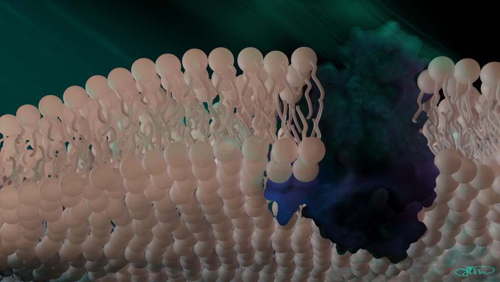
 They found that there are four phospolipid molecules that signal to the body rest of the body that the cell they are around is damaged and needs to be properly taken care of. A phospholipid is a molecule with phosphate head with a lipid tail, it is commonly found throught out the body. Phospholipids are the molecules that make up the majority of cell membranes, the picture above is depiction of a cell membrane. A lead member of the team Professor Kagan says, "Scientists have long known that these lipids were important for encasing the cell and giving it structure... What they didn't know is that they do so much more, including communicating and signaling messages like 'danger' inside the cell itself, to other cells and to the cellular community as a whole, so that organisms can function in a coordinated way." (UPMC). Their findings have also suggested that ferroptosis can be used to treat the underlying cell that cause radiation sickness. Althought apoptosis has already been decoded every controlled cellular death pathway is different and every new decoded pathway can help uncover why certain pathways are not working. Certain pathways are also used in certain types of cells, apoptosis is not the path choosen everytime in every sitituation. Figuring out what pathway is used at what time and to what cells are the focus on is the next medical breakthrough that will help treat and cure many terrrible dieases.


Helmholtz Zentrum Muenchen - German Research Centre for Environmental Health. (2016, November 15). Cancer research: How cells die by ferroptosis. ScienceDaily. Retrieved November 16, 2016 from www.sciencedaily.com/releases/2016/11/161115083915.htm

 University of Pittsburgh Medical Center (UPMC). (2016, November 14). International team decodes cellular death signals. ScienceDaily. Retrieved November 15, 2016 from www.sciencedaily.com/releases/2016/11/161114110708.htm

Thursday, April 14, 2016

The Common Flu Could Change Everything for the Treatment of Cancer


Based off of research recently done at Queen Mary University of London, a common flu virus will be used in the near future to help patients overcome resistance to certain cancer drugs and improve the way that these drugs kill cancer cells. What is known is that viruses can be altered to specifically enter cancer cells and rapidly multiply until the cell bursts. After the cell bursts, the virus's copies then spread to surrounding cancer cells and the process repeats all over again leaving healthy cells unharmed. The virus can never reach every cancer cell before being killed off by the body's immune system and this is where the problem lies.

 In Pancreatic Cancer specifically, there is a drug that patients with this specific type of the disease take called Gemcitabine. Gemcitabine causes the DNA in cancer cells to be damaged and therefore cannot divide successfully. A process called apoptosis is triggered which causes unhealthy and damaged cells to self-destruct. Gemcitabine works perfectly at treated and reversing cancer, however after a short amount of time the cancer cells learn to delay apoptosis in order to repair the damaged DNA. Because of this, the cells survive and begin to divide and spread again which counteracts the medication.

Under the work of Dr. Gunnel Hallden at Queen Mary University of London Barts Cancer Institute, a new genetic modification to a virus called Adenovirus, making it a stronger enemy against cancer cells. Dr. Hallden explains that by switching off a specific gene in the virus that counteracts apoptosis, cancer cells infected with the virus can no longer delay apoptosis meaning that they die before dividing. Although it does not kill every single cancer cell, it works nicely with anti-cancer drugs to kill more cancer cells and lowers the risk of cancer cells becoming resistant to apoptosis.

Dr. Hallden states that "many cancers, including Pancreatic cancer, become resistant to treatments like Gemcitabine and currently there is no way to get around that. However, the virus that we have modified re-sensitizes the resistant cancer cell by preventing the cell from repairing itself. The virus alone will kill some tumor cells but in combination with the drug, the number of cells that are killed is greatly increased."  She also points out that because the virus improves the efficiency of the drug, less of it may have to be given and therefore less resistance to the drug will occur.

Further research has to be done as Dr. Hallden's work is in the early stages but what we can expect to see next is other versions of the adenovirus to better understand how it enhances cell killing. The team strongly believes that they have found a very promising route to treating and curing Pancreatic cancer and with that will leads to more promising routes for treating and curing all types of cancer.

I find it so interesting to read that we coming so far along in treating and curing cancer. When you think about cancer from a genetic point of view, it seems rather simple to notice that the way to stop cancer from reproducing and spreading is to damage the DNA in the cancer cells which prevents them from reproducing. This would ultimately halt cancer in its tracks. I also found it very interesting and did not know about apoptosis at all. I had no clue that there was a process in cells are programmed to die. I look forward to following the work done with Pancreatic cancer and hope that they find a cure very soon as though cancer of the Pancreas is known to be one of the worst kinds.


Thursday, December 4, 2014

Innate immune system condemns weak cells to their death

Link to the Article


Molecular Biologist at the University of Zurich and Columbia University have demonstrated that the innate immune system plays a key role in this mechanism. Cancer cells however can also use this to cause cells that are important for healthy tissues to die. The scientists used a fruit fly model to show that during cell competition, programmed cell death is activated in the weaker cells. This cell death is caused by Spätzle, a signaling protein that docks onto toll related receptors. These toll receptors normally trigger a defensive reaction to bacterial or fungal reactions, but can also trigger apoptosis in weak cells. However, sometimes the stronger cell is not a healthy cell such as a tumor that can outcompete the weaker cells. Then healthy cells fall behind in fitness and undergo apoptosis.

This research is important for cancer researchers and can help contribute to early detection cancer. The innate immune system can help identify faster growing cells that are not yet malignant cells. This presents a new way to fight cancer at an earlier stage and help treat it before the patient is terminal.


Survival of the Fittest Cell

Survival of the fittest doesn’t only happen in the wild. It also happens in the body. When people think of apoptosis, many think of a cell that is sick or damaged that must be removal. However, it has been discovered that if there are weak cells surrounded by healthier cells, these weak cells will be killed off. This is a problem when the stronger cells are cancer cells. Molecular biologists from University of Zurich and University of Columbia have been the first to publish that the only way for this process to happen is when one has an innate immune system.


The biologists proved their theory with an experiment with fruit flies. They saw that apoptosis was induced by “Spätzle”, which is a signaling protein that binds to the Toll-related receptors. This was known information, however; before this experiment, it was thought that this protein only reacted to the presence of foreign bacteria and fungus. It is now known that it also responds to less fit cells. The only part the team is not sure about whether it is the weak cell that triggers the binding or the stronger cells around it. As Professor Konrad Basler says, “We still don’t know whether this involves the voluntary or forced suicide of the less-fit cells.”


While this may sound like a good thing. Weaker cells are being conquered by healthier, stronger cells who are in better shape to do the work it needs to. However, what happens when the stronger cells are not good cells. This cell activity is often found in cancerous tissue. This raises big problems. However, it gives bigger hope to cancer research. Scientists hope that this discovery might lead to early detection of the disease, possibly before the cells are even malignant.

http://www.sciencedaily.com/releases/2014/12/141204140648.htm

Sunday, November 23, 2014

3D Model of Bax Protein Newly Proposed!


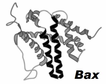
The protein Bax is one of great interest to scientist. Bax is one of the proteins responsible for cell death by puncturing holes in the mitochondria of cells. This in turn causes proteins in the organelle to leak into the cytoplasm of the cell, resulting in cell death through apoptosis. Which some may believe this protein to be troublesome, cell death is sometimes needed. Especially in the situation of cancer patients. Cancer is the over multiplication of cells, if there was a way to use this Bax protein against cancer cells, it would greatly revolutionize cancer treatments. The only problem was that until very recently, scientists did not know what Bax exactly looked like. They knew the crystal structure of the truncated core of the domain, but not the full length structure on the protein.


However, scientists at the Freie Universität Berlin, the University of Tubingen, and the Swiss Federal Institute of Technology in Zurich have developed a 3D model of the Bax protein. With this model, scientists now know how the protein looks and acts. The most significant discovery was that the hairpin at helices 5-6 of the protein form into a clamp-like dimeric structure. This structure is thought to be the mechanism that punctures the mitochondria. The punctures are actually formed by the structure pinching the membrane bilayer. One scientist, Enrica Bordignon, explains how important this discovery is by saying, “If we understand how the Bax protein deforms the membrane and forms pores, we are one step closer to understanding how cells die, and thus one step closer to the development of new cancer drugs which should promote cell death.” If the Bax protein could be better understood and could be used to control the apoptosis of cancer cells, it would be an incredible advancement in the field of cancer treatment research.

Sunday, November 2, 2014

Significant Research Breakthrough for Lupus, Cancers and Neurodegenerative Diseases

Research teams from the Walter and Eliza Hall Institute combined to discover both the three-dimensional structure of a key cell death protein, Bak, as well as an understanding of the first steps in how Bak causes cell death. Apoptosis, programmed cell death, occurs when chemical signals activate Bak and Bax. The activation of Bak and Bax tells the cell to die by targeting the mitochondria. When apoptosis is working effectively unwanted cells are killed and removed from the body. However, when trouble with apoptosis occurs harmful cells, such as cancer cells, continue to grow or healthy cells die unnecessarily, such as what happens in Alzheimer's disease.


Specifically, the team’s research showed how Bak changes from one form to another form in order to initiate apoptosis. Additionally, the research team discovered how the Bak and Bax protein dimers attach to the mitochondria and perforate the mitochondria. It has been understood that the Bak and Bax dimers open the mitochondrial surface; however, the mechanism for how the dimers perforate the mitochondria was previously poorly understood.


Scientists originally believed that Bak and Bax poked a hole through the surface of the mitochondria; however, the team’s research disproved this belief. Instead the team discovered that the dimers attach onto the oily surface of the mitochondria and crowd the surface until holes form in the surface. What still remains unknown is exactly how the proteins come together to destroy the mitochondria and initiate apoptosis.


I found this article very interesting because this small development will lead to a much greater understanding of apoptosis in the future. Now that researchers are able to understand the structures of the proteins involved apoptosis they have the necessary information to begin advancing towards a deeper understanding of apoptosis, specifically the initiation of the process. This research will be crucial to understanding how to regulate apoptosis to combat diseases such as lupus, cancers and neurodegenerative diseases.  

Sunday, March 30, 2014

Neighboring Cells Alerted To Protect Themselves By Dying Cells In Fruit Fly

When a cell's DNA becomes irreversibly damaged, it goes through a process called apoptosis, or self-destructs so the damaged DNA does not replicate. TinTin Su Ph.D., and her collaborators at the University of Colorado, found that dying Drosophila melanogaster larvae cells alert neighboring cells that they could also die. They tested this by using ionized radiation (IR) to activate apoptosis in the wing imaginal disc cells, or the premature form of the fly's wings. What they found was that the neighboring cells responded by activating bantam which made it more difficult to kill by IR. They determined that receptor tyrosine kinase was behind this and the dying cells turned the receptor on. Dr. Su believes that if this protective mechanism also works in mammals, it could affect the results of using cytotoxic agents and radiation in cancer therapy.
This discovery is very interesting. The next step is to figure out if this does actually happen in mammals. It is important to figure this out because if it in fact does occur, it would make radiation for cancer much more difficult to work.


Original article: http://www.medicalnewstoday.com/releases/274633.php
Related article: http://science.howstuffworks.com/life/cellular-microscopic/apoptosis.htm

Thursday, November 21, 2013

Cancer Suppressing Gene

A new gene has been revealed by Adelaide researchers to play an important role in suppressing lymphoma.  Lymphoma is a type of cancer that starts out in immune system cells when they are in a mode of uncontrollable growth and multiplication, which eventually leads to a tumor.  The gene discovered to supress lymphoma is the caspase-2 gene, which is related to a family of proteins that are essential for the self destruction of cells.  This gene was first discovered by Professor Sharad Kumar, and for the past twenty years his laboratory has been researching the processes by which cells committ suicide.  Cell death is vital in order to maintain the correct number of cells in the body and to delete cells that are potentially harmful.  Kumar states, "Cell death and survival are controlled by a large number of genes, and aberrations in these genes are often linked to diseases. For example, an inability for cells to evade apoptosis is a well-known hallmark of cancer".  The caspase-2 gene could actually prevent tumor formation by making cells predisposed to cancer maintain the right number of chromosomes.  Kumar adds, "This research not only provides new information on the development of cancer, it also defines how caspase-2 can potentially work as a tumour suppressor gene".  This article was very interesting to be because it has the possibility to lead to significant progress in cancer research and treatment.  Although research is still being done it is a nice shot in the arm towards treating this disease.

File:Reed-Sternberg lymphocyte nci-vol-7172-300.jpg


Friday, November 23, 2012

Designed genetic circuit may force cancer cells to commit suicide

According to MIT News , synthetic biologists from MIT and ETH Zurich have designed a genetic circuit that programs cells to detect cancerous cells and force them to commit suicide, while still keeping healthy cells unharmed. This genetic circuit is programmed to make a "decision based on multiple inputs." In other words, this genetic circuit is made of genes programmed to detect molecules that are specific to a particular cervical cancer that these researchers have chosen to work with, HeLa cells.   According to Popular Science, this genetic circuit contains genes that could identify up to "five cancer-specific molecules and their concentrations." Once cancerous cells are detected, the genes from this designed circuit would release protein that would provoke the cells to commit suicide, or apoptosis (cell death).  For the cells to undergo apoptosis, the five "characteristics" must be present.

The researchers that took part in this project designed this genetic circuit by studying a type of newly discovered genetic material known as microRNA and choosing that to be their "target."  MicroRNA, according to MIT News, is a snippet of RNA that helps regulate gene expression by selectively destroys messenger RNA. The reason why these researchers chose microRNA was because a large amount of particular types were found in cancer cells. According to the article, each different kind of cancer contained its own microRNA profile.

The microRNA profile of the cervical cancer the researchers chose to experiment with, HeLa, contained six microRNAs that were able to be identified. They were found in large quantities as well as unique to this particular cancer.  They then created a synthetic gene that would code for a protein that would trigger apoptosis, hBax. This gene could be turned off by high levels of microRNA that are usually found low in HeLa as well as by low levels of microRNA that are usually found in large amounts in HeLa. However, this research is still fairly new and still being tested. According to these researchers, this process needs to be tested on living animals. While this system detects up to five characteristics in a cancer cell, they are still working on trying to get the system to identify more markers.

It is one step closer to finding another form of cancer treatment. Despite the fact it is still in its early stages and is not readily available to be used as a form of treatment just yet, it is still amazing that these researchers are able to design and program a gene to do this, while still keeping healthy cells unharmed. If this were to be a readily form of cancer treatment, would it be able to detect pre-cancerous cells rather than just cells that are already in their cancerous state? With further study and experimentation, I believe that they will be able to revise the design so that the circuit can detect cancerous cells earlier on and use this system for other kinds of cancer, not just this particular cervical cancer.