Showing posts with label fibroblasts. Show all posts

Friday, November 24, 2023

Neuroimmune Crosstalk Role in Heart Tissue Repair

Researchers at the Max Delbrück Center have discovered that zebrafish regenerate heart tissue using communication signals between their nervous and immune systems. In general, myocardial infarctions happen when blood vessels supply blood and nutrients to the heart, resulting in portions of the afflicted heart tissue dying. Since humans are unable to grow new heart cells to reduce the damage, they instead form scar tissue that weakens the pumping power of the heart overtime. Unfortunately, even stem cell research has been proven to be unsuccessful here.

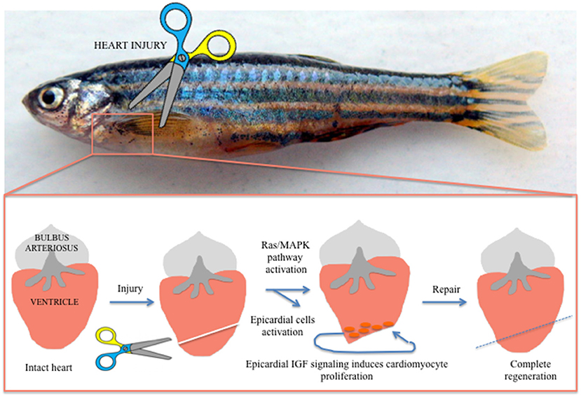
Interestingly enough, signals between the autonomic nervous system (ANS) and immune system were found to play pivotal roles in scarring cand tissue regeneration. To observe this communication, researchers induced an injury into the hearts and several macrophage receptors of zebrafish larvae. After noticing that ANS adrenergic signals resulted in macrophages multiply and regenerating heart muscle, the research team genetically engineered the fish larvae so that the signal couldn’t enter the macrophage cell. The research study found that interrupting the adrenergic ANS signal deactivated the macrophages and induced heart scarring. In difference, when macrophages are activated by these signals, they communicate with fibroblasts and promote regeneration at the damaged site, creating an environment conducive for the growth development and growth of blood, lymph, and heart vessels.


A rather interesting study, this research’s findings provide an insight into how the regeneration of human heart muscle tissue can be made foreseeable. By better understanding the differences in signaling between zebrafish and humans, biologists can better understand why cardiac tissue does not regenerate, find methods to navigate a path to initiating the regeneration process, and even how to better treat heart attack patients’ conditions.

For more information, the news article has been linked here and the published journal article has been linked here.

Tuesday, December 15, 2015

The regeneration of fibroblasts might just save your heart

Dr. Srivastava and his team began reprogramming fibroblasts, or scar- forming cells, in heart muscles of live mice. Fibroblasts secrete extracellular matrix required for the formation of connective tissue and other fibers. These heart muscle fibers had once been damaged by heart attacks. Heart attacks cause these fibers to become encased in scar tissue and cause cells in that area to stop beating. A new treatment has involved a “cocktail of genes” in order to regenerate or reverse the damage done by myocardial infarction. 
Dr. Srivastava and his colleagues also began injecting the same therapy of three genes into fibroblasts in foetal heart cells, embryonic stem cells, and neonatal skin cells in petri dishes. Dr. Srivastava reported low numbers of transformed cells but at least a partial transformation of 20 % were capable of transmitting electrical signals.

By using a combination of genes the doctors have managed to turn human fibroblasts into heart cells that conduct electrical signals in lab petri dishes. This is an incredible feat. The ability to take a muscle that was once destroyed and restore it for a better function is just remarkable. Although the success is low at the moment this just brings scientists a step closer in the practice of cardiovascular medicine. I really thought this article to be positive. Just knowing we are a bit closer at helping people with heart disease is really fantastic.  

Saturday, November 8, 2014

Using Fibroblasts to Create New Blood Vessels

        Cardiovascular researchers at Houston Methodist have learned that they can use fibroblast cells and convert them into endothelial cells which will create blood vessels. Fibroblasts are the cells that cause scar tissue and are abundant in the human body. John Cooke, the study's main researcher, says that this is the first time that small molecules and proteins have been converted into a therapeutic cell type.  Cook's hope for this discovery is that it will be used to improve the healing of cardiovascular injuries and other injuries throughout the body that require an increase in circulation.There have already been studies done that use viruses to transform cells into those need in the body, but there are limitations and many risks that go along with this form of transformation. It is believed that using small molecules and proteins will be more safe for use.
The top picture shows fibroblasts stained blue, and the bottom picture shows the amount of fibroblasts that transformed into endothelial cells after treatment with poly I:C and VEGF

         The new method and Cook and his other researchers proposed involves exposing the fibroblasts to poly I:C (polyinosinic:polycytidylic acid) that will cause the cells to think that they are being attacked by a virus. Poly I:C is a small segment of RNA that binds to the host cell receptor TLR3. This viral attack caused the fibroblast cells to reorganize their nuclear chromatin, which allowed genes that had previously been blocked off to be expressed. Factors, such as VEGF, were the applied to the fibroblasts because these factors are known to cause certain cells to convert into endothelial cells. This treatment caused 2% of all the fibroblasts in the body to be transformed into endothelial cells, the same percentage outcome as using a virus to transform the cells. Cooke claims that he has unpublished work that shows that up to 15% of the fibroblasts can be converted using his method.
       
         In order to prove the effectiveness of the new cells, Cooke injected the cells into mice that had the need for new blood vessels in their hind limbs for circulation. Once the cells were introduced to the mice, the blood vessel number increased in the hind limbs and blood flow was improved. Cooke believes that his findings will pave the way for more studies to continue and possibly lead to finding ways to regenerate mass amounts of damaged tissue in humans.

       This is a very interesting study that goes to show that there are many different ways to manipulate the cells of the human body, and as our knowledge of genetics continues to grow, many more interesting findings like this will occur.

Original Article: Reprogrammed cells grow into new blood vessels

Friday, November 7, 2014

New Blood Vessels from Reprogrammed Fibroblasts


Scar cells(top) transformed into blood vessels. The
proof of transformation is indicated by the red color in
the bottom picture. The red is an indicator for CD31,
a protein made by blood vessels. 

Cardiovascular scientists from Houston Methodist, Stanford University, and Cincinnati Children’s Hospital teamed up in a joint effort to study fibroblasts, cells that cause scarring. Our bodies are filled with an immense amount of fibroblast. Through their study, the scientists discovered that the fibroblasts can be transformed into endothelium, a cell type that forms the lining of blood vessels. The method first involves polyinosinic:polycytidylic acid (poly I:C), a segment of double-stranded RNA, being introduced to fibroblasts. Poly I:C binds to TLR3(toll-like receptor 3), which fools the fibroblast cell into believing it was attacked by a virus. This resulted in a rearrangement of nuclear chromatin, which allowed genes to be expressed that were once restricted. After rearrangement, the fibroblast was treated with VEGF, Vascular endothelial growth factor, which allowed the fibroblasts to become endothelial cells.

"To our knowledge, this is the first time that trans-differentiation to a therapeutic cell type has been accomplished with a small molecules and proteins," explained chairperson, John Cooke, M.D. Houston Methodist Research Institute Department of Cardiovascular Sciences.

The next step in their research involved taking the transformed fibroblasts and introducing them to immune-deficient mice. The immune-deficient mice had poor blood circulation, however, with the transformed fibroblasts the number of vessels in the limbs of the mice increased, and ultimately improving circulation.

"The cells spontaneously form new blood vessels -- they self assemble," Cooke said. "Our transformed cells appear to form capillaries in vivo that join with the existing vessels in the animal, as we saw mouse red blood cells inside the vessels composed of human cells."

Although procedures like this have been performed, this is the first time a small molecule has been reprogrammed.  Research groups were able to generate endothelial cells from infectious viruses, viruses that were programmed to manipulate DNA cells. However, this process involves a more complicated approach. Viruses also have the potential to damage patient’s chromosomes. The small-molecule transformation of cells is a safer approach that will be utilized in clinical trials. The new research also helps our society take one step further into regenerative medicine. The new discovery will definitely help humans who suffer from poor blood circulation and cardiovascular health affects, by improving their condition through the formation of new blood vessels.

Article Related: Fibroblasts - http://ghr.nlm.nih.gov/glossary=fibroblast

Thursday, April 18, 2013

Turning Skin Cells into Functioning Brain Cells

The following is an amazing discovery described in the ScienceDaily. Researchers at the Case Western Reserve School of Medicine have actually found a way to turn skin cells into functioning brain cells. This is help many patients whose brain cells get destroyed with conditions such as multiple sclerosis, cerebral palsy, and other myelin disorders. The technique involves coverting fibroblasts, which are present in most organs and skin, into oligodendrocytes, which are responsible for myelinating the neurons of the brain by manipulating proteins. They call is cellular reprogramming. The initial study used mouse cells, and their next step is to see how safe it is for humans. This finding a real breakthrough and can be used to treat a variety of genetic myelin disorders. There is not much other information on the study because it has only been done on mice. They get closer and close to making it safe for humans everyday.


[caption id="attachment_8004" align="aligncenter" width="448" caption="Neurons in the Brain "]Neurons in the Brain [/caption]


I think this is an astounding discovery that deserves great merit. It would help so many patients if and when it is available as a plausible treatment for myelin degeneration. When further studies are done in the field I would love to read about it, and I hope for the best.