Showing posts with label skin cells. Show all posts

Wednesday, May 6, 2026

Using Bioinformatics to Investigate Keloid


Image: Jiao H, Zhang T, Fan J, and Xiao R (2017). Keloid Dermis at Different Depths.


A recent study investigated the biological mechanisms behind keloid formation and may help lead to improved methods for preventing and treating the condition. Keloids are a type of fibrotic skin disorder caused by excessive growth of scar tissue, often resulting in raised scars, pain, and limited movement. Although keloids are common, the exact causes of their development are still not fully understood. Current treatment options are also limited in effectiveness.

Researchers used data from the Gene Expression Omnibus (GEO) database to identify genes that are expressed differently in keloid tissue compared to normal tissue. Bioinformatics analyses were performed to examine gene enrichment patterns as well as cell and tissue specific expression.

The analysis identified the gene Homeobox D8 (HOXD8) as a major contributor to keloid development. HOXD8 expression was found to be approximately two times higher in keloid tissues than in normal tissues. When researchers suppressed HOXD8 activity, the growth of keloid fibroblasts was significantly reduced, while cell death increased. These effects were also linked to changes in the Wnt/β-catenin signaling pathway.

Overall, the study suggests that HOXD8 plays an important role in promoting keloid formation through its effects on signaling pathways in keloid fibroblasts. These findings provide new insight into the molecular basis of keloid disease and indicate that HOXD8 could serve as a promising target for future therapies.


link:
https://www.sciencedirect.com/science/article/pii/S1687850726002268


additional:
https://www.frontiersin.org/journals/physiology/articles/10.3389/fphys.2017.00885/full

Sunday, November 26, 2017

Transgenic skin received by Syrian refugee

A Syrian boy with a rare genetic disease has received an almost entirely new epidermal layer of skin from his own stem cells. This boy has a disease called Junctional epidermolysis bullosa  and has a mutated gene causing the basement membrane of the containing laminin 332 does not develop properly. so a group of scientists and pediatricians created Karatinocyte cultures from the boys stem cells , they used invivo and invitrio renewment of human epidermal cells, the boy will still have to live with the disease for the rest of his life and may also have issues with other systems in his body this disease is also known to affect the mucosa. besides the amazing accomplishment that this undergoing was, researchers were also able to end  a long standing discussion whether the epidermis was regenerated by many equally powerful progenitor cells or a fewer number of individual stem cells. The later of the two is true according to the researcher.

I believe any advancement in stem cell research is a good thing , the more we as a society can create new cells from a persons original cells is amazing and could help such a young child have anew lease on life from the cultivation of his own stem cells is remarkable.


Link 1

Thursday, October 12, 2017

Monkeys with Parkinson's disease benefit from human stem cells


A article on science daily was published regarding how monkeys with Parkinson's disease are benefiting from human stem cells.
Parkinson's disease degenerates cells in the brain known as dopaminergic (DA) neurons. When the symptoms of Parkinson's are found, it most likely appears that more than half of the DA neurons are lost in the brain of the individual. Neurosurgeons are planning on using DA neurons created from iPS cells to treat these patients with the disease. iPS cells are made from blood and skin cells which makes them easily obtainable. This idea is being tested for its safety and reliability, so they are trying the experiment on monkeys that have Parkinson's by transplanting the new cells in the monkeys brains.

I believe this could be a remarkable and cheap breakthrough if it works. Scientists were usually using fetal tissue to create DA neurons . Studies mentioned on how the transplant of these cells aid and help relieve symptoms of the disease. But using fetal tissues in general is a controversial topic. The use of iPS cells can help substitute the use of fetal tissue to treat patients.


https://www.sciencedaily.com/releases/2017/08/170830132224.htm

http://www.nature.com/nature/journal/v548/n7669/full/nature23664.html?foxtrotcallback=true

Friday, November 18, 2016

Why wounds heal more slowly with age

For years now, people have seen obvious evidence that supports the fact that older individuals do not heal as rapidly as younger individuals. Children have been known to be more resilient because of their hope, and young, strong body systems that are constantly overcoming obstacles and our elders are treated with delicate care and caution. Aging occasionally triggers various inheritable diseases or mutations that are caused by genetics. It has been a mystery as to what is causing the body to start slowing down the healing process as we age, so researchers at Rockefeller University began to explore the correlation between ages.

Healing wounds, whether superficial or extremely severe, is one of the bodies most complex processes because it involves various cell types, signaling systems, and molecular pathways that have to communicate and with time, close in and once again form its protective barrier. Immune cells and skin cells both work together to form a scab and keratinocytes act as a sheet and pile in beneath the wound.


Lab mice were used as the model organism for the experiment at the University. They were separated into two groups: 2 month-old mice and 24 month-old mice, which is the equivalent of having groups of 20 year-olds and 70 year-old humans. Researchers observed molecular changes in the mice's skin and it was found that the keratinocytes in the older mice moved much slower to their place below the scab, than those of the younger mice. This lack of speed in the keratinocytes resulted in the wounds taking days longer to heal in the older mice than the younger mice.

Researchers learned that after an injury, the keratinocytes communicate with specialized immune skills that reside in the skin. They produce proteins called Skints that are responsible for keeping the immune cells around, and together they help in filling in the gap of the wound. The scientists thought that they could boost Skint signaling in older individuals and speed up healing times. So they used a protein released by immune cells after an injury. Using petri dishes, scientists applied the protein with skin tissue samples from both young and old mice to discover that they had enhanced keratinocytes' migration, and the older keratinocytes more closely resembled those of the younger mice.
This study provides hope for the future in terms of speeding up the healing process for older individuals. It is only normal for healing speeds to begin to lag after a certain age, but if scientists are able to produce a drug that would be capable of counteracting that, like they think is possible, it would minimize people's pain and discomfort greatly, as well as increase health.

https://www.sciencedaily.com/releases/2016/11/161117151059.htm

Monday, March 28, 2016

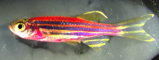
Skinbows leave no pot of gold but they do shine!

        Today I've brought you a look into a topic we've covered in class and in lab. Fluorescence in bacteria is one thing, but the study conducted in Nature magazine shows viewers what happens when you add these fluorescent proteins into zebrafish DNA. Scientists have studied how DNA can be transferred into other organisms and through experimentation have found that when the skin cells regenerate in the fish after sustaining damage they actually made several different bundles of color. When these pigments are allowed to be varied with these colors rather than following the normal genetic make-up, the expression is pretty interesting to see. It doesn't help the fish to be fluorescent in the wild as they can be spotted by predators. But it gives us a way to view what happens when skin cells are doing their designated function, as well as explains a little as to why scarred tissue appears different than the skin around it.
        There is a video of the what the cells look like during the process of regeneration in the link above to the magazine. And I have included a link to the research paper here. Other than these pictures they have plenty of other data that goes into detail about the experiment itself. They talked about barcoding the superficial epithelial cells, (SEC's) those on the outer part of the skin, so that each variation of color is linked to specific strains of cells. They documented the interactions the cells underwent to achieve these interactions.

       Perhaps research like this can lead to advances in skincare. If we know in which concentrations the variations of our own skin cells are affected when we get scarred or burned perhaps we can learn how to improve skincare treatments for lacerated and burn victims. Or at least find safer ways to get the right kind of tan for the summer. There are many possibilities to explore.

Monday, October 19, 2015

Creating "Old" Cells from Skin Cells

In the article scientists create 'old' cells  the author discussed how skin cells can now be converted into neurons without losing their "age".  This was done by Prof. Gage and colleagues, who decided to use the direct conversion technique to try (and succeed) in creating neurons out of skin cells, rather than the traditional induced pluripotent stem cell method, which was previously (and currently is) done to do the same conversion.
The problem scientist had with the pluripotent method, although it worked, was that this reset epigenetic signatures of the old cell and made them like that of a younger neuron.  This can become a problem when it is used to look at diseases such as Alzheimer's disease and other age related neurological diseases.  This new technique is and can be very important, because by using the new method of conversion scientists are able to bypass the stem cell precursor state, and as a result create an "induced neuron".

I find this to be an amazing advancement, because as they mentioned in the article this allows researchers to identified age related changes within the brain.  Which with the induced pluripotent stem cell method was more difficult because after going through that process they would end up with young neurons, which did not seem to have the same qualities as older neurons.

Thursday, September 10, 2015

Scientists map 1,000 molecular tools common to cells across tree of life


One of the largest experiments ever undertaken by seven research groups in three different countries into animal molecular biology has found that across the tree of life many organisms share protein machines that help cells to function. The tests were done on a range of organisms from slime mold, yeast, worms, to sea urchins, sea anemones and flies, mice, and even humans. The new study identified instructions for almost 1,000 protein complexes that were found across the tree of life.

I think this article is interesting because scientist have found over the years many ways that different organisms or similar and different but not too many ways that all organisms are linked. Knowing that we all share proteins for cell function can hopefully progress different studies in science.


Related article (This research can be applied to many studies)


Thursday, May 1, 2014

Study turns skin tissue from infertile men into early-stage sperm cells

Scientists have currently turned skin tissue from infertile men into early-stage spermcells in a study that raises hopes for new therapies for the condition. It is said that about 1% of men cannot make any sperm, a condition known as azoospermia, while a fifth of men have low sperm counts
Live sperm seen through a microscope. The success of this was actually unexpected and has surprised many scientists. Most scientists thought that it was impossible for infertile men to make any sperm, but this breakthrough has defied those odds. Most of the men that took part in the study had many genetic defects on their Y sex chromosome which meant that they could not produce healthy sperm. In this study researchers took skin cells from three infertile men and converted them into stem cells, which can actually grow into almost any tissue in the human body.
Cyril Ramathal of Stanford University said, "What we found was that cells from men who did not possess sperm at the time of clinical observation were able to produce the precursors for sperm.”
The study found that the skin cells from the infertile men grew into fewer early-stage sperm cells than cells taken from normally fertile men. Even though this research has just started scientist suspect that the converted skin cells might have grown into mature sperm cells if they had been transplanted into the infertile men's testes. If this suspicion is confirmed then it would be possible to restore male fertility by taking the skin cells of men and turning them into stem cells.
This is a great discovery and I think that the research for this should continue. Infertility in men seems to be growing and is a tragic thing. So, this study will definitely help those men that are completely infertile.

Thursday, April 10, 2014

Britain's first cloned dog is born – and described as 'ridiculous waste of money'

This article discusses a woman that had won a contest that allowed her to get her family dog cloned with the charge covered by the company. The price tag for this experiment was 60,000 pounds and being described by scientists as a "ridiculous waste of money". The cloning was successful and a new puppy was born but the dog will not be the same as the beloved animal it was cloned from. The family pet was a 12 year old dachshund and scientists took skin cells which they used to implant in an egg and into a surrogate dog. This idea is really a true bust because what people don’t understand is that the dog has almost no chance of being the “same” dog as the one it was cloned from. The dog may look the same but the emotions of the dog will never be exactly the same; it’s possible the dog will be completely different. There are as a lot of ethical problems with this idea due to the fact that most cloned dogs do end up dying. In a different study one out of 1,095 puppies cloned survived; many dying within three weeks or before birth.

Woman holding puppy Mini-Winnie


The idea of this is nice for people that want to bring their beloved animals that they have lost back into their family; but sadly there are too many holes in this for it to really be practical. The idea of bringing home a beloved animal look alike and have it be nothing like the original almost seems like it will cause more harm than good. It seems like it is much better to give a sheltered animal a chance and have it become a new family member rather than trying to bring back an old one; it would also save tens of thousands of dollars. This article gives a list of animals that have been cloned.

Thursday, December 1, 2011

Skin Cells Detect UV light


In a recent article, biologists from Brown University have reported that our skin cells can detect ultraviolet light.  This UV light is detected from a receptor within our eye known as a photosensitive receptor. During this new study , resarchers were able to detect a new rhodopsin receptors which are known to detect UVA light when the melanocyte skin cells fluoresce when calcium level hit a high level after a large exposure of UV light. During these experiments, researchers noticed that when they reduced rhodopsin levels, the calcium levels then decreased and when they increased these levels, the calcium levels increased as well. Our skins DNA tries to protect us from sunlight damage, although after a heavy amount of it, the damage will still occur. Even though scientists learned these possible way to check for how in fact our skin cells react to sunlight, we still need to protect our skin from the sunlight even though we love to tan.  Sunscreen is still the safest way to tolerate sunlight.