Showing posts with label cell membrane. Show all posts

Monday, November 25, 2019

A Step Toward Putting Batteries in Humans


Ryota Iino is a Japanese researcher with the Institute for Molecular Science of the National Institutes of Natural Sciences. He and the other researchers focused on the V1 rotational motor found in the membrane of healthy cells. The motor acts as a pump, moving sodium ions through the cell membrane. They used an advanced probe to observe a single molecule from the bacteria Enterococcus hirae (above). They used the high-resolution images to develop an understanding of how the motor turns chemical energy into mechanical energy. They found that it works closely with another motor complex called Vo to convert the energy of the sodium ions into electrochemical potential energy similarly to how electric eels generate electric energy.

With further research, Ryota Iino plans to further understand the complexity of the energy conversion. If they are able to learn enough about the motor complex, he believes it could be possible to design an implant that could be put in electric eels of even humans to generate energy.


Tuesday, December 1, 2015

Scientists Identify Two Genes that ‘Shut Down’ HIV-1 Virus


An international group of researchers has uncovered the mechanisms of genes that may inhibit the virility of the HIV-1 virus. The two proteins, SERINC5 and SERINC3 are typically found in  the cell membrane of a cell. The HIV-1 virus has 9 genes, one of them coding for the protein HIV-1 Nef, which accounts for much of the viral ability of HIV. HIV-1 Nef also prevents SERINC5 and SERINC3 from reaching the cell membrane or being incorporated into new virions. In the absence of Nef, new virions can leave the host cell and attempt unsuccessfully to infect other cells because the SERINC proteins prevent the HIV virus from leaving the viral envelope. With the presence of SERINC proteins, research say the virility of HIV is reduced by 100-fold.

The research is pointing towards a promising future in viral studies, as HIV has become rampant over the years. Current medicine usually keep the virus at bay and infected individuals manage to have normal, functioning lives. Even so, they still have to live with the stigma of their infection and infected people who do not get diagnosed early may suffer greater consequences. With SERINC's anti-retroviral ability, this could lead to a more effective treatment option for HIV/AIDs and treatment of other enveloped viruses.

Original article

SERINC

HIV/AIDS

Sunday, November 2, 2014

Cell Division Without Cells

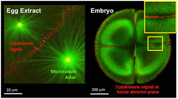
Cell division is the essential process of life. The final step in cell division, when the two daughter cells split from each other by a process known as cytokinesis, has fascinated scientists for a long time, mainly because of how difficult it is to find cells undergoing this process. To this day, it is still a mystery as to how a cell can signal where the furrow should be. This is such a problem because of how difficult it is to see and test such a process in living cells. 

Harvard Medical School systems biologists report that they have "reconstituted" cytokinesis "complete with signals that direct molecular traffic" without the cell. The scientists combined frog-egg extracts with lipid membranes that have the ability to mimic the membrane of the cell. They essentially built a "cell-free" system that summarizes how a cleavage furrow is made. This system has two huge advantages: it makes the furrow-building events easier to see by expanding the scale, and it also gives the researchers an easier way to manipulate the proteins involved in the process. They can now more easily remove and return proteins to see how they affect the process of cytokinesis. 

The main problem with this is that cytokinesis is completely dependent on having a membrane to furrow. By having a "cell-free" system, the membrane must be removed. This experiment is only possible by having a controlled, flat membrane. This membrane is made by having two layers of artificial lipid supported by glass. 

The process begins by crushing unfertilized egg cells from the Xenopus frog and isolating their internal contents. The scientists built artificial centrosomes and fluorescent microtubules and then mimicked fertilization. They then used labeled proteins to "visualize the self-organization of structures required for the cell division process." This process uses fluorescence microscopy in ways that are not possible with living cells in tissue cultures. The final part of the process was the model of the cell membrane. 

"To really prove that we reconstituted the cytokinesis signal, we needed to add the bilayer membrane and then see if it could recruit the proteins that would be on the cortex of the cell.... That's the signal to the membrane we were looking for," said Christine Field, HMS instructor in systems biology. Field had used actin in their system which was used to organize the long filaments and meshwork and allowed the components to move and change shape, including the membranes and other cellular components. Only when the actin is around can the microtubules signal to the plasma membrane and also to the actin cortex that forms on top of the plasma membrane. It was found that multiple types of signaling complexes are at work in the cleavage furrow. They "talk" from the microtubules to the actin cortex and then to the cell membrane. 


"The beauty of this system is that we have reconstructed cytokinesis from its individual structural components: actin, microtubules and membranes," said Aaron Groen, a researcher in systems biology. "And we can now begin to spatially manipulate these components, which is not possible in live cells. I'm very excited by the possibilities."

Although their work is the most basic aspect of science, cell division is the center of embryonic development, stem cell renewal and cancer. The understanding of this process is relevant to many types of diseases. 

It was exciting to see such an incredible breakthrough in science today. This can be the beginning to finding cures for countless types of diseases and I am very interested in where this research team is going next with their studies. I won't be surprised at all if this process is what helps in future medical breakthroughs.


Article: http://www.sciencedaily.com/releases/2014/10/141031133545.htm 

Related Article: http://hms.harvard.edu/news/cell-division-minus-cells 

Monday, November 26, 2012

New DNA Nanotech Mimics Membrane Channels in Cells

[caption id="attachment_6345" align="alignleft" width="250" caption="This nanodevice is formed by 54 double-helical DNA domains on a honeycomb lattice."][/caption]

Physicists from Technische Universitaet Muenchen and the University of Michigan have shown how to construct synthetic membrane channels from the use of DNA nanotechnology. The DNA based channel is shaped after natural membrane channels in lipid bilayer membranes. "Membrane channels are tube-like structures made of proteins, which pierce the barriers and regulate the two-way exchange of material and information between the inside and outside."-Medical News Today. The device's stem is 42 nanometers long and has a internal diameter of two nanometers. The stem is partly protected by a barrel-like cap that is surrounded by cholestorol units which enables it to bind to the lipid membrane of a cell. Once attached to a cell wall, the stem sticks through and forms a pore. The pores formed from the synthetic membrane channels have electrical conductivity just like ion channels in natural cells and might be able to act like voltage controlled gates. The transmembrane current could be adjusted by changing structural details of the synthetic channels and geometric and chemical tailoring of the membrane channels can also be accomplished by this technology.

This little DNA based device could be used as molecular sensors, antimicrobial agents, and drivers of novel nanodevices. "If you want, for example, to inject something into a cell, you have to find a way to punch a hole into the cell membrane, and this device can do that, at least with model cell membranes," says TUM Prof. Hendrik Dietz, from TUM Institute for Advanced Study. This nanotechnology is also propused to imitate the action of viruses or phages by breaking through the cell walls of targeted bacteria to kill them, used as nano-needles to inject material into cells, and basic studies of cell metabolism. Dietz even said "We might be able to mimic natural ion pumps, transport proteins, and rotary motors like the enzyme responsible for synthesizing ATP".

This artical is excting and marks a new step in the development of nanotechnology. This device created by the Technische Universitaet Muenchen and the University of Michigan has opened many doors to the inside of a cell. In the future I think this device will be able to help prevent and treat illnesses.