Harvard Medical School systems biologists report that they have "reconstituted" cytokinesis "complete with signals that direct molecular traffic" without the cell. The scientists combined frog-egg extracts with lipid membranes that have the ability to mimic the membrane of the cell. They essentially built a "cell-free" system that summarizes how a cleavage furrow is made. This system has two huge advantages: it makes the furrow-building events easier to see by expanding the scale, and it also gives the researchers an easier way to manipulate the proteins involved in the process. They can now more easily remove and return proteins to see how they affect the process of cytokinesis.
The main problem with this is that cytokinesis is completely dependent on having a membrane to furrow. By having a "cell-free" system, the membrane must be removed. This experiment is only possible by having a controlled, flat membrane. This membrane is made by having two layers of artificial lipid supported by glass.

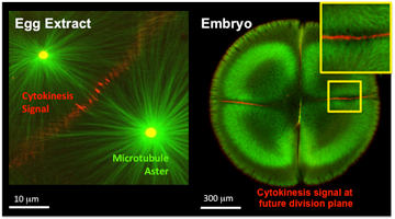
The process begins by crushing unfertilized egg cells from the Xenopus frog and isolating their internal contents. The scientists built artificial centrosomes and fluorescent microtubules and then mimicked fertilization. They then used labeled proteins to "visualize the self-organization of structures required for the cell division process." This process uses fluorescence microscopy in ways that are not possible with living cells in tissue cultures. The final part of the process was the model of the cell membrane.
"To really prove that we reconstituted the cytokinesis signal, we needed to add the bilayer membrane and then see if it could recruit the proteins that would be on the cortex of the cell.... That's the signal to the membrane we were looking for," said Christine Field, HMS instructor in systems biology. Field had used actin in their system which was used to organize the long filaments and meshwork and allowed the components to move and change shape, including the membranes and other cellular components. Only when the actin is around can the microtubules signal to the plasma membrane and also to the actin cortex that forms on top of the plasma membrane. It was found that multiple types of signaling complexes are at work in the cleavage furrow. They "talk" from the microtubules to the actin cortex and then to the cell membrane.
"The beauty of this system is that we have reconstructed cytokinesis from its individual structural components: actin, microtubules and membranes," said Aaron Groen, a researcher in systems biology. "And we can now begin to spatially manipulate these components, which is not possible in live cells. I'm very excited by the possibilities."
Although their work is the most basic aspect of science, cell division is the center of embryonic development, stem cell renewal and cancer. The understanding of this process is relevant to many types of diseases.
It was exciting to see such an incredible breakthrough in science today. This can be the beginning to finding cures for countless types of diseases and I am very interested in where this research team is going next with their studies. I won't be surprised at all if this process is what helps in future medical breakthroughs.
Article: http://www.sciencedaily.com/releases/2014/10/141031133545.htm
Related Article: http://hms.harvard.edu/news/cell-division-minus-cells
No comments:
Post a Comment